Keywords
Forensic Anthropology, CT Images, Sternum, Sex determination

This article is included in the Manipal Academy of Higher Education gateway.
Forensic Anthropology, CT Images, Sternum, Sex determination
We sincerely thank the reviewer for the detailed and constructive comments, which have significantly improved the quality of our manuscript. In response to the major concerns, the statistical analysis was revised by replacing the univariate analysis with a multivariable logistic regression model that included age as a covariate to address potential confounding. The revised model identified combined length (CL) and manubrial width (MW) as independent predictors of sex, with model adequacy confirmed using the Hosmer–Lemeshow goodness-of-fit test. To improve the clinical and forensic applicability of the findings, the ROC analysis was expanded to include optimal cut-off values, sensitivity, specificity, the Youden Index, and 95% confidence intervals. The interpretation of the Sternal Index (SI) was also corrected to reflect reverse-direction discrimination, indicating that higher SI values are more characteristic of females. The effect of age was further clarified, demonstrating that it was not an independent predictor after adjustment. Although observer reliability analysis was not performed, this has now been acknowledged as a limitation, and future studies have been recommended to include intra- and inter-observer reliability assessments using the Intraclass Correlation Coefficient (ICC). To enhance reproducibility, the Materials and Methods section has been expanded to provide detailed CT acquisition parameters, scanner specifications, reconstruction settings, and image analysis software details. The Data Availability Statement has been revised to clarify that data sharing is restricted by ethical and institutional regulations but may be considered upon reasonable request and appropriate approvals. Statistical reporting has been strengthened through the inclusion of confidence intervals, standardized reporting of software versions, p-values, and decimal precision. Finally, all minor issues, including typographical errors, repetitive text, and overstatement of conclusions, have been addressed.
See the authors' detailed response to the review by Yasin Etli
Sex determination is one of the important aspects in forensic anthropology for establishing the biological profile of unidentified deceased individuals, as it helps in narrowing the possible identities for individual identification.1 Although the pelvis and the skull are the most dependable skeletal remains for sex determination, often these bones are not available due to decomposition, fragmentation, or severe damage in mass disaster cases.2 In such scenarios, an alternative flat bone, the sternum, due to its robust nature, as it is positioned centrally within the thoracic cage, is well preserved and is the most resistant bone that can recover, even in adverse conditions.3 Several sternal morphometric studies have shown significant sexual dimorphism in sternal measurements, with males having larger dimensions than females.1,2 Across different populations, ML, CL, and MSL have been demonstrated to be reliable parameters to determine sex among various parameters studied.1,4
Traditional Morphometric methods for sex determination are limited by preservation, observer bias, and direct handling of skeletal remains. CT has overcome these limitations by being a reproducible, non-invasive, and accurate 3D analysis of the bones5 to determine sex without direct contact and is more reliable than traditional methods.1,6
Several studies have explained that morphometric standards are population-specific, as bone growth and development are influenced by various factors such as environment, genetics, and nutritional factors.1,2,7 Several studies on different populations, global as well as in India, have consistently emphasized that a regression model applied to one population cannot be applied to another population.8–10 Several CT-based sternal studies have been conducted on various populations; a specific morphometric standard for the Karnataka population is yet to be explored. Hence, the current study aims to establish sexual dimorphism of the sternum using CT images and to determine the most reliable parameter(s) of the sternum for accurately determining sex in the Karnataka population, which may be used as future reference data for forensic anthropology and medico-legal practice.
Sex determination is a primary step in forensic identification, as it greatly narrows the range of possible identities and plays a major role in subsequent biological profiling in the field of forensic anthropology, and helps in medico-legal investigations. Edwards et al.,11 and Zech et al.,12 pointed out the difficulty associated with sex determination in mass disasters, explosions, fire accidents, mutilation, and in cases of high degrees of decomposition. Although DNA examination is one of the most precise methods for determining sex, Battan et al.6 emphasized that “degraded samples, contamination, cost, and technical limitations often limit the use of DNA analysis”; in such situations, forensic anthropology is an important field that can be used to determine sex from skeletons.
Traditionally, the pelvic bones and the skull are the most reliable, as these bones show the highest degree of sexual dimorphism, as noted by Franklin et al.2 and Zech et al.12 A study on the South Indian population conducted by Nagesh et al.13 showed that the acetabulum-pubis index accurately identified 83% males and 81% females. This study also highlighted that skeletal indices vary among populations and cannot be generalized. Likewise, a study conducted in the Greek population showed that sex classification varied from 79.7% to 95.4% depending on the index employed.14 In a Turkish population, Gülhan15 showed that 3D pelvic indices obtained from CT scans are as reliable as conventional osteometry. A CT-based study conducted on the North Indian population showed strong sexual dimorphism in the coxal, ischiopubic, and sacral indices.16 Though the pelvic gridle is considered to be the most dependable skeletal part for sex determination because of its strong sexual dimorphism, its practicality in forensic studies is often limited because the pelvis is often broken, incomplete, or absent in cases of trauma, mass disasters, and advanced decomposition.17 Thus, the assessment of other skeletal remains is required to ensure accurate sex determination in the absence of a pelvis.
Skull- the commonly used skeletal remains for sex determination, followed by the pelvis. A skull-based study conducted in the South Indian population found that biauricular breadth was the most reliable parameter among the 26 chosen parameters, with an accuracy of 85.7%.18 In another study on the isolated mandible conducted by Sharma et al in the Indian population showed that even though diagonal length & minimum ramus breadth were statistically significant, overall sex determination was only 60%.19 In a study on the Turkish population, using 3D CT, Dereli et al.20 stated that the glabella was found to be the most influential trait with 92% accuracy, but the female skull showed lower classification accuracy compared with the male skull. Likewise, Zaafrane et al.21 established craniometric standards using CT scans in a modern Tunisian population, achieving up to 90% accuracy with multivariate logistic regression models, underscoring the importance of population-specific equations. However, despite the enhanced accuracy and reproducibility associated with the use of CT-based methods, sex determination from the skull is limited by its dependence on multiple landmarks and statistical analyses, which make it less accurate than the pelvis, especially when it comes to fragmented specimens.20,21
Rahmani et al.22 measured the patella dimensions by MRI in an Iranian population and reached a maximum accuracy of 85.7% using transverse and craniocaudal measurements. Liu et al.23 assessed sphenoid sinus morphometry by MRI in Chinese adults and found an accuracy of 63.3%, indicating fair discriminatory ability and underlining the ability of MRI to image deep structures without superimposition of bones. More recently, Cavlak et al.24 used convolutional neural networks on MRI slices of the patella and achieved a maximum accuracy of 88.88%, reflecting the increasing use of artificial intelligence in forensic anthropology.
While MRI has the advantage of not using radiation and the ability to study living individuals, some drawbacks are mentioned: reliance on the machine settings, high cost, soft tissue artifacts, lack of availability in forensic practice, and only moderate accuracy in certain areas.23,24
In a metric study of the long bones in a contemporary Japanese population, Sakaue25 analyzed 47 variables for the humerus, ulna, radius, femur, and tibia using discriminant function analysis and obtained sex discrimination accuracies of 91–95%. The analysis also showed that epiphyseal breadth measurements, especially the distal articular width of the humerus and the proximal epiphyseal breadth of the tibia, were more effective than bone lengths. The presence of sexual dimorphism was observed to be more evident on joint surfaces, whereas measurements of shafts and lengths were relatively less reliable. However, the author stressed the importance of population specificity, and different regressions must be used on different populations. Six femoral measurements were assessed by Mall et al.26 in a modern German population, with classification accuracy of up to 91.7% for a combination of midshaft diameter and head circumference, with transverse head diameter being the single best predictor. Likewise, Steyn & Işcan27 studied the femoral and tibial dimensions of South African whites and concluded that the distal epiphyseal breadths were the most sexually dimorphic features, with accuracies of 86–91%.
Studies have shown consistently that the use of breadth and epiphyseal measurements is a better discriminant than the use of length alone. However, it was emphasized by all authors that discriminant functions cannot be generalized over different ethnic groups and that population specificity is required.26 Amer et al.28 analyzed Egyptian subjects by combining the parameters from MRI of hand and foot bones and obtained a cross-validated accuracy of 90.2% by multivariate discriminant analysis, indicating that the combination of parameters improves the accuracy. Although long bones offer a high degree of accuracy when complete, their recovery may be restricted in the case of fragmented specimens or epiphyseal damage.25
However, to overcome the limitations of the traditional skeletal components, there have been several studies on flat and irregular bones, like the scapula,3,29,30 clavicle,30 vertebrae,31 ribs,32 and sternum.4 These bones are often retained even in adverse forensic situations. Of these, the sternum, an isolated bone, is one of the important bones in the determination of sex because of its robust nature and centrality to the thoracic cage.33
Another aspect of the sternum is that, compared to other bones, it is more likely to be available even when the bones are fragmented.34 The presence of sexual dimorphism in the sternum was proven by Uysal Ramadan et al.4 who established that the sternum is effective in determining sex even in the absence of other bones. The effectiveness of the sternum was further proven by Changani et al.9 in the Gujarati population. A study conducted by Koçak et al.32 demonstrated that the superior-inferior height of the sternal end of the rib was the most accurate feature in Turkish, with accuracies of 86.5–88.6%. A study established that there are sex differences in the sternum, reflecting its efficacy in providing the necessary information for forensic analysis.9
As forensic radiology is increasingly used in forensic science, computed tomography has been recognized as a non-invasive, precise, and repeatable technique in the skeletal analysis of individuals.29 CT-based sternal analysis has shown high accuracy in sex determination, with discriminant function models showing classification accuracy rates above 80% and as high as 95.5% when multiple variables are combined, thus emphasizing its ruggedness for forensic applications.35 Zhang et al.1 have confirmed that it is a reliable technique for sex determination without the need to physically touch the bone. Rodríguez et al.5 have highlighted the importance of avoiding observer bias by utilising computed tomography in forensic anthropology in their study. Ali et al.36 studied an Egyptian population and demonstrated the importance of MDCT in estimating sternal parameters, which are essential for logistic regression-based sex-estimation models.
While MRI provides high contrast for soft tissues, it is technically demanding for bone morphometry due to the low proton density and poor signal of cortical bone, thus making it less ideal than CT for detailed analysis of bony morphology.37 Moreover, CT scanning enables accurate reconstruction of complex bone geometry that aids in landmark identification and measurement, which is important in sex determination, where minute dimensional differences are employed.
Thus, when compared with the conventional morphological, manual osteometry, and MRI-based methods, the CT scan method stands out as a better technique for sex determination because of its non-destructive, three-dimensional, and reproducible nature, and the ability to be applied to both dead and living individuals, hence justifying its choice as the chief methodological approach in this study.
Different studies have used various sternal parameters for sex determination. A study by Zhang et al.1 in the western Chinese population measured the lengths of the manubrium, the width of the manubrium, the sternal body length, the combined sternum length, and the sternal index. In their study, significant sexual dimorphism existed in all the measured lengths, and a classification accuracy of about 80% using multivariate CT-based analysis. Combined sternum length and dimensions of corpus sterni were used by Franklin et al.2 in the Australian population, resulting in an accuracy above 80%.
Earlier, in the Turkish population, Uysal Ramadan et al.4 found that the parameters of mesosternal length and combined length were highly reliable. Another study conducted in the Turkish population by Koşar et al.8 found that the combined length and total length of the sternum alone were sufficient to predict sex with close to 85% accuracy. More parameters were introduced by Ghorbanlou et al.,38 such as the sternum area, which were found to have an accuracy rate above 90% in the Iranian population.
A study conducted on the African population by Banyeh et al.7 found that most linear parameters of the sternum were significantly larger in males, whereas the sternal index was smaller for males. However, it was emphasized that the model results should be generalized with caution, considering the variability among populations.
Indian studies have considerably enhanced regional forensic standards, although the sample population remains restricted. On one hand, a study by Changani et al.,9 undertaken among the Gujarati population, found that the best predictor for sex determination was the sum of the lengths of the manubrium and sternum, while the sternal index showed limited predictive value. Meanwhile, the sexual dimorphism of the sternum was again re-established by Chowdhuri et al.39 among the Eastern Indian population, which revealed moderate accuracy in determining sex based on sternal morphometric parameters. More recently, Sravan et al.10 established that the highest predictive potential was offered by the measurement of the mesosternal length among the Central Indian population. For all these populations, it has, however, been evident that the sternal index has low forensic accuracy.
Studies conducted by Changani et al.9 among the Gujarat population, Ali et al.36 among the Egyptian population, and Banyeh et al.7 among the Ghanaian population have concluded that females exhibit higher sternal index values. In contrast, all other parameters were consistently higher in males, which led to a considerable overlap between the sternal index value for both males and females, making it an unreliable parameter for sex determination.
From the existing literature, it is evident that morphometric standards differ greatly from population to population. Zhang et al.,1 Franklin et al.,2 Rodríguez et al.,5 Banyeh et al.,7 Koşar et al.,8 and Ghorbanlou et al.38 stress this important fact: do not apply these equations to other populations, as genetics, environment, nutritional, has great impact on the shape of the human skeleton. This fact is particularly relevant given that the country of our interest, India, has rich geographical and ethnic diversity.
The parameters chosen in the current study- Manubrium Width (MW), Combined Length (CL), and individual Sternebra widths (S1W to S4W)- have been well-established by prior research for sex determination.1,9,10 The values for ML, MW, MSL, and CL have constantly exhibited significant dimorphism between the sexes worldwide, as well as within Indian individuals.1,4,8,10 The inclusion of individual Sternebra widths has been justified by previous investigations indicating their contributory role when combined with other parameters, even though these parameters have been reported to yield lower accuracy.7,38
Population specificity is one of the essential aspects in forensic sex determination studies, since bone morphology is affected by genetic, environmental, nutritional, and socio-cultural variables. Because of the great ethnic diversity of the Indian population, skeletal standards based on Western, Central, or Northern Indian populations may not necessarily represent morphometric differences among the Karnataka population. Thus, by examining CT-based sternal morphometry among a specific South Indian population, the current study fills an important gap in the forensic literature and improves the applicability of sex determination in medico-legal practice in the Karnataka region.
This retrospective study included 146 sternal CT images (73 males and 73 females) from the Karnataka population, obtained from a medical institution in Karnataka.
The research included participants aged 25 to 80 years whose sternal CT images had sufficient image quality for morphometric measurements. Participants below the age of 25 years, above 80 years, CT scans of the sternum with trauma, deformities, or poor image resolution were excluded. Ethical clearance (IEC No. IEC2:483/2025) was obtained from the Kasturba Medical College and Hospital Ethics Committee. As per the Institution Ethics Committee (IEC), it is mandated to get consent from the Medical Superintendent of Kasturba Medical College to access the CT images, and written consent with participant ID was obtained to access the participant’s CT images. CT imaging was obtained using a 128-slice Incisive CT scan machine (Philips Health Care Suzhou Co., Ltd., China). Imaging was obtained with a thickness of 3 to 5 mm, while the reconstructions were made in the coronal orientation using the B-reconstruction filter. Measurement of the sternum was done with InstaRISPACS version 5.0.0.
The following measurements were taken into consideration for morphometry and recorded in millimeters:
1. Manubrium width (MW)- The greatest transverse distance between the lateral margins of the manubrium at their widest point.1
2. Combined length (CL): Sum of the manubrium and sternal body lengths.4
3. Sternebra 1 Width (S1W)- The maximum transverse width of the first sternebra of the mesosternum, measured at its greatest width.2,7,10,36
4. Sternebra 2 Width (S2W)- The maximum transverse width of the second sternebra of the mesosternum, measured at its greatest width.10
5. Sternebra 3 Width (S3W)- The maximum transverse width of the third sternebra of the mesosternum, measured at its greatest width.2,7,10,36
6. Sternebra 4 Width (S4W)- The maximum transverse width of the fourth sternebra of the mesosternum, measured at its greatest width.10
7. Sternal Index (SI)- SI = {Manubrium Length}/ {Mesosternal Length} * 100.10
The data was analyzed using SPSS/version 31 for the continuous variables. Mean, Standard Deviation, and median (IQR) were calculated. For the categorical variable, frequency- percentage was used to calculate. Student’s t-test was used for independent samples to compare males and females. The value of P(<0.05) was considered statistically significant. Multivariable logistic regression was used to identify the most reliable parameter(s) for sex determination.
Table 1 shows that the study included 146 sternum CT scans, with 73 males and 73 females ( Table 1).
| Frequency | Percent | ||
|---|---|---|---|
| Sex | Male | 73 | 50.0 |
| Female | 73 | 50.0 | |
| Total | 146 | 100.0 | |
Table 2 shows that all sternal parameters exhibited statistically significant dimorphism, with CL and MW greater in males (p < 0.001) than in females. Among them, CL showed the greatest sex difference with a mean of (132.3 ± 8.62) in males and (114.4 ± 8.48) in females, indicating the overall larger sternal size in males ( Table 2).
The S1W to S4W were also found to be significantly greater in males compared to those of females. Statistically significant differences were found for S1W (p < 0.001), S2W (p = 0.003), S3W (p = 0.013), and S4W (p < 0.001), indicating sexual dimorphism for the transverse dimensions of the sternebra. In contrast, the SI of the females in the study population was observed to be significantly higher (51.47 ± 8.52) compared to that of males (48.26 ± 6.25), with p = 0.010 ( Table 2).
Table 3 presents the multivariable logistic regression analysis after adjustment for age. Among the evaluated sternal parameters, combined length (CL) remained the strongest independent predictor of sex (OR = 1.222, 95% CI: 1.128–1.323; p < 0.001), followed by manubrium width (MW) (OR = 1.166, 95% CI: 1.033–1.317; p = 0.013). Age, sternebral widths (S1W–S4W), and the sternal index (SI) were not independently associated with sex after adjustment (all p > 0.05). The Hosmer–Lemeshow goodness-of-fit test indicated that the model demonstrated an adequate fit (Table 3).
Table 4 shows the receiver operating characteristic (ROC) analysis for the morphometric parameters of the sternum for estimating sex. From all the parameters, CL was found to have the highest discriminatory power with an AUC of 0.926 (95% CI: 0.884–0.968; p < 0.001) and excellent accuracy. The parameter MW had an excellent discriminatory power with an AUC of 0.850 (95% CI: 0.788–0.911; p < 0.001). The measures of sternebral widths (S1W–S4W) showed fair discriminatory power with AUC values between 0.607 and 0.665. SI had an AUC of 0.393 (95% CI: 0.301–0.485; p = 0.022), indicating that the parameter is reverse discriminative since its values were greater in females than in males (Table 4) and the Figure 1 shows the ROC curve analysis (Figure 1).
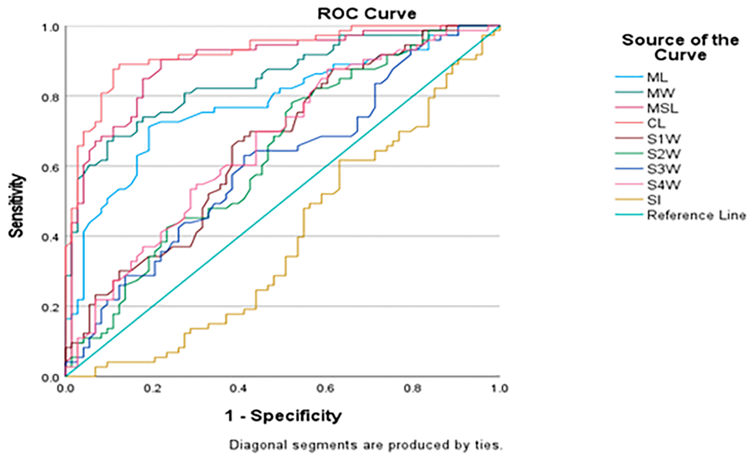
Table 5 summarizes the optimum cut-off values determined by the ROC curve analysis for the assessed morphometric features of the sternum. From all the measures taken, the combined length (CL) exhibited the highest discriminative power, with an optimum cut-off value of 121.20 mm, with 90.4% sensitivity, 79.5% specificity, and the highest Youden Index (0.699). The second best was manubrium width (MW), which had an optimum cut-off value of 57.05 mm, with 68.5% sensitivity, 89.0% specificity, and a Youden Index of 0.575. Sternebral width exhibited relatively poor discriminative power, with Youden Indices of 0.178 to 0.274.
Sex determination forms the most fundamental step in establishing the biological identity of unidentified human remains. In scenarios where the most sexually dimorphic pelvis and skulls are unavailable, the sternum can serve as an alternative skeletal remain due to its anatomical position and resistant nature.2,4,36
All the assessed parameters showed significant sexual dimorphism, with males having higher values than those of females (p < 0.05), except for SI, which was found to be higher in females (p = 0.010). A statistically significant difference in the mean age of the males (46.56 ± 12.69 years) and that of the females (51.93 ± 13.04 years) was observed (p = 0.013). Even though an age difference between males and females was found to be significant, age was still used as a covariate within multivariable logistic regression to account for its potential confounding effect. However, age was not considered the principal variable of interest, as the sternum usually finalizes its growth by early adulthood and maintains stable linear dimensions.1,2 Among all the parameters, the CL of the sternum was found to have a statistically significant difference between the sexes (p = <0.001), with males having a higher mean value (132.3 ± 8.62) than females (114.4 ± 8.48), while the SI remained higher in females (51.4729 ± 8.52) than in males (48.2642 ± 6.25). These findings are consistent with several studies among different populations.1,2,9,10,40,41
The consistent observation of larger sternal dimensions in males across various populations was explained by biological factors that are related to sexual dimorphism in bone development. During puberty, increased androgen levels influence bone formation on the periosteum and the development of the musculoskeletal system in males, leading to a larger and stronger thoracic cage.1,2 Additionally, the greater muscle mass in males puts more stress on the thoracic cage, which contributes to the growth of the sternum.
Logistic regression with multivariable analysis was carried out with adjustment for age to determine the independent predictors of sex. Combined length came out as the most significant independent predictor (OR = 1.222, p < 0.001), while manubrium width was the second (OR = 1.166, p = 0.013). Age, sternebral widths, and sternal index were not significantly associated with sex. The goodness-of-fit of the regression model was confirmed using the Hosmer-Lemeshow test (p = 0.963).
According to ROC analysis, the discriminatory power was highest for the combined length (AUC = 0.926) and then manubrium width (AUC = 0.850). The sternebral widths were moderately discriminative, while the sternal index was discriminating in the reverse direction-discrimination (AUC = 0.393). This result is further supported by various other studies where combined sternal length is effective in sex determination.1,2,4,8,9
The higher discriminatory power of CL may be attributed to the cumulative contribution of both manubrial length and mesosternal length. Sexual differences tend to be better explained by cumulative measurement than by individual parameters.42
However, in some studies, there was a variation in predictive parameters among different populations7,10 which could be due to the differences in body morphology among different populations, genetics, nutrition, environmental factors, and body size factors.38
This highlights that though sexual dimorphism is evident in the sternum among different populations, the parameters may vary.
The present study’s results demonstrated the potential of CT-based sternal morphometry in the field of forensic anthropology for determining sex. A high degree of discriminatory accuracy for CL with AUC = 0.926 and the optimal cut-off value of 121.20 mm yielded 90.4% sensitivity and 79.5% specificity, indicating excellent diagnostic performance study, making this parameter reliable for determining sex in the Karnataka population. Moreover, CT imaging is a non-destructive method, providing a clearer view of the skeletal structures and the possibility of using existing data.5
The current study, a retrospective morphometric analysis using CT scans, examined the sexual dimorphism of the sternum in the Karnataka population and successfully developed population-specific reference values for sex estimation. All the linear dimensions showed significant sexual dimorphism, with CL being the most reliable parameter, followed by MW, while the SI was found to be in the reverse direction-discrimination.
Thus, the current study reiterates that CT-based sternal morphometry is a non-destructive and reproducible technique for sex estimation. The results form the basis for future multicentric studies with larger sample sizes, uniformly distributed age groups, and employing more advanced statistical tools. Future studies incorporating external validation and assessment of intra- and inter-observer reliability would further strengthen the applicability of CT-based sternal morphometry.
Ethical clearance (IEC No. IEC2:483/2025) was obtained from the Kasturba Medical College and Hospital Ethics Committee.
The research was done according to the requirements of the Institutional Ethics Committee (IEC). As per the IEC process, the committee mandates the researcher to get consent to access the CT images from the Medical Superintendent of Kasturba Medical College, and written consent with participant ID was obtained to access the participant’s CT images. Thus, consent was obtained from the Medical Superintendent before collecting the data.
| Views | Downloads | |
|---|---|---|
| F1000Research | - | - |
|
PubMed Central
Data from PMC are received and updated monthly.
|
- | - |
Provide sufficient details of any financial or non-financial competing interests to enable users to assess whether your comments might lead a reasonable person to question your impartiality. Consider the following examples, but note that this is not an exhaustive list:
Sign up for content alerts and receive a weekly or monthly email with all newly published articles
Already registered? Sign in
The email address should be the one you originally registered with F1000.
You registered with F1000 via Google, so we cannot reset your password.
To sign in, please click here.
If you still need help with your Google account password, please click here.
You registered with F1000 via Facebook, so we cannot reset your password.
To sign in, please click here.
If you still need help with your Facebook account password, please click here.
If your email address is registered with us, we will email you instructions to reset your password.
If you think you should have received this email but it has not arrived, please check your spam filters and/or contact for further assistance.

Comments on this article Comments (0)