Keywords
Amblyopia, occlusion therapy, age, lazy eye
This article is included in the Eye Health gateway.
Amblyopia, occlusion therapy, age, lazy eye
Amblyopia, (‘blunt vision’ in Ancient Greek), also known as lazy eye1, is a visual deficiency in an eye that is otherwise physically normal, or that is greater than would be expected from any structural abnormalities of the eye. It is thought that amblyopia results from inadequate stimulation of the fovea or peripheral retina and/or abnormal binocular interaction, resulting in different visual input from the foveae2. It has been estimated to affect 2–5% of the population3. The global prevalence of amblyopia has not significantly changed over the years4.
Amblyopia results in the loss of binocular vision, which is manifested as absent stereoscopic depth perception, poor spatial acuity, low contrast sensitivity and reduced sensitivity to motion5. This can be detected clinically by assessing whether the patient has difficulty seeing three-dimensional images on autostereograms6.
Amblyopic patients can have poor spatial acuity, low contrast sensitivity, and reduced sensitivity to motion5. They may also have poor binocular vision and limited depth perception which can be detected on autostereograms6.
The largest study on patient response to amblyopia therapy to date was conducted by the Pediatric Eye Disease Investigator Group (PEDIG) in the USA7, which reported that amblyopia due to strabismus and/or anisometropia responds similarly to part-time occlusion therapy in children between the ages of three and seven years. PEDIG also reported that about 75% of children with previously untreated amblyopia respond to treatment with only two or more lines of improvement on the Early Treatment Diabetic Retinopathy Study (ETDRS) chart up to 18 years of age8. The upper age limit for optimal therapy was considered to be in the range of nine to ten years9–11. The American Academy of Ophthalmology recommends that all children be considered for treatment of amblyopia, regardless of age12.
Recent findings in neuroplasticity have replaced the formerly-held position that the brain is a physiologically static organ and have shown that it changes throughout life13. Much evidence of neuroplasticity has been found in adults14,15. According to Levi and Polat, significant improvement of Vernier acuity in adult amblyopes occurred following stereoacuity by using 3-D video games14. Similar findings were announced by researchers of the Goldschleger Eye Research Institute, Tel Aviv University16. The mean improvement in distance and near acuity in amblyopic eyes by 12 months was 3.3 and 1.9 lines log MAR (minimum angle of resolution) respectively.
In 2009, a study conducted by MK Mallah and coworkers at Queen's University17, Royal Victoria Hospital, Belfast, Ireland, showed that older people (60–80 years) with a history of amblyopia who develop visual loss in the previously normal eye can experience recovery of visual function in the amblyopic eye over a 12 month period. This recovery in visual function occurred following visual loss in the other, previously normal, eye; distance improved to 3.3 lines and near to 1.9 lines log MAR. This improvement appeared to be sustained.
There are unsubstantiated beliefs that it is more difficult to treat amblyopia in older age groups, that children that have already received failed amblyopia therapy do not respond to treatment and that full-time occlusion therapy may result in occlusion (disuse) amblyopia of the good eye. The aim of this study was to assess whether these beliefs are true.
This was a prospective trial of 115 consecutive cases with unilateral, severe amblyopia conducted at a tertiary referral center in Lahore, Pakistan, from January 2010 to October 2012 after obtaining ethical committee approval by the University of Health Sciences. Eligibility criteria included an age of over three years, visual acuity in the amblyopic eye from 6/60 Snellen’s or even counting fingers only, visual acuity in the sound eye of 6/6, an inter eye acuity difference of three or more lines, and the presence or history of an amblyogenic factor such as refractive, deprivational and strabismic components that met study-specified criteria for strabismus and/or anisometropia. A complete ophthalmic examination was performed by only one ophthalmologist (SI). This included examining the fixation pattern of both eyes, presence or absence of a phoria or a tropia by a cover-uncover test, fundus examination and color vision using Ishihara color plates. Any case with an organic cause for visual loss was excluded from the study after a thorough ophthalmological examination. Assessment of visual acuity of either eye for both near and distance acuity (Snellen’s in literate patients and Kays picture chart for illiterate children (3–5 year olds)), refraction and Best Corrected Visual Acuity (BCVA) was performed by a trained optician who was masked to the study and patient’s demographics.
There were 59 female and 56 male patients and the cases were divided into three age groups:
Group A: age 3–7 years (mean age 5.38±2, median 5 yrs) = 38 cases (37.25% of the total number).
Group B: 8–12 years (mean age 9.73±1.73, median 10 yrs = 41 cases (40.20% of the total number).
Group C: 13–35 years (mean age = 19.26±6, median 17 yrs = 36 cases (22.55% of the total number).
All patients had some degree of anisometropia; two children had stimulus deprivation amblyopia due to traumatic cataract (aged five and seven years) and 82 patients presented with strabismus.
All cases were prescribed an optimal refractive correction for a month after which full-time occlusion therapy was started. After verbal informed consent was obtained from either the parents or the guardians, patients were provided with stick-on commercial eye patches and were instructed to wear the patch over the good eye as soon as possible after waking up in the morning. They were strictly instructed not to take it off during the day, only when they were about to sleep at night. Patients were instructed regarding the near visual activities they should perform for 3–4 hours/day. All patients and both of their parents or care-takers were thoroughly counseled about regular follow up, compliance towards the patching schedule and engaging the children towards near visual activities. The latter included coloring, drawing, reading large prints initially and then as near vision improved, shifting to smaller prints, playing video games on their personal computers and hand held video games inter alia.
Patients were closely followed-up at regular intervals of one day/year age; for instance four year olds were checked every fifth day and six year olds, every seventh day. Children more than 7 years old were followed up every two weeks. Patients were instructed to come into the clinic wearing the occlusive patch. First the vision of the amblyopic eye was checked and then that of the occluded eye; children were instructed to wear the patch immediately thereafter. Any change in fixation pattern of the two eyes was noted after removing the patch.
The criteria for a successful therapeutic outcome in this study was regarded as a maximum visual recovery (achieving 6/6 Snellen’s). Occlusion therapy was continued until this was achieved in all age groups. If after two months of full-time patching no visual improvement was noted, further therapy was stopped. If therapy was successful it was gradually reduced over the next seven weeks and then stopped. The weaning protocol adopted was one day off in the first week, two days off the second week, three days off the third week and so on until the seventh week, when occlusion therapy was discontinued.
Children in the 3–7 year age group were followed-up weekly after therapy discontinuation and any drop in visual acuity during weaning was monitored. Patients over the age of seven were followed-up after every two weeks until occlusion therapy was successfully finished in the seventh week.
Patients were followed-up at two weeks, one month, two months and then every three months for the next 18 months after stopping full-time occlusion.
The clinical demographics of the patients are summarized in Table 1. Success was defined as equalization of visual acuity in both eyes, i.e. 6/6 (Snellen’s).
| Person specific characteristics | No. |
|---|---|
| Total patient number | 115 |
| Age: 3–7 years | 38 |
| Age: 8–12 years | 41 |
| Age: 13–35 years | 36 |
| Gender, female (%) | 59 |
| Gender, male (%) | 56 |
| Traumatic cataract | 02 |
| Strabismus | 82 |
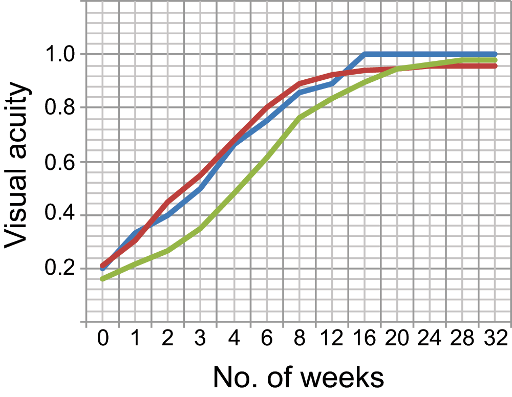
Improvement in visual acuity noted with full-time occlusion therapy was 6/6 Snellen’s in 38/38 (100% success) in group A. In group B, 38 cases out of 41 achieved 6/6 vision (92.68% success). In group C, 35 cases out of 36 achieved 6/6 (97.22%) success (Table 2). The average time duration for successful amblyopia therapy in group A was 8 ± 1 weeks, in group B was 9 ± 2 weeks and in group C was16 ± 2 weeks (Figure 1).
| Age group | Patients achieving 6/6 | % | Patients not achieving 6/6 |
|---|---|---|---|
| 3–7 years | 38/38 | 100 | 0/30 |
| 8–12 years | 38/41 | 92.68 | 3/41 = 7.32% |
| 13–35 years | 35/36 | 97.22 | 1/36 = 2.88% |

The blue line represents group A (3–7 years); red represents group B (8–12 years); green represents group C (13–35 years). Visual acuity axes are a conversion of Snellen’s scores to numerical values: 6/60–0.1; 6/36–0.167; 6/24–0.25; 6/18–0.333; 6/12–0.5; 6/9–0.667; 6/6–1.
Visual acuity improved even in the "unsuccessful cases" (not achieving 6/6). The three unsuccessful cases in group B improved from 6/60 to 6/12 and the one unsuccessful case in group C was found to be due to eccentric fixation. Hence an improvement of five lines did occur even with eccentric fixation.
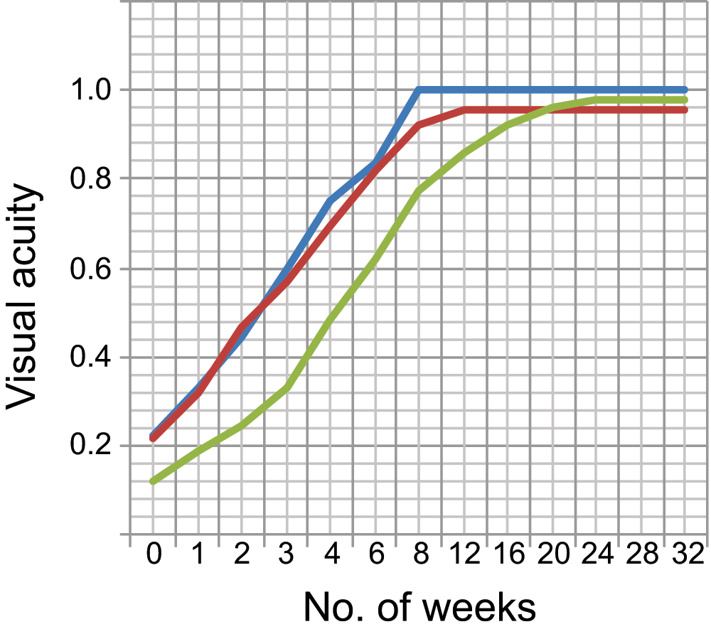
Patient compliance to therapy was found to be an important factor influencing clinical improvement in vision in this study. Groups A and C were noted to be more compliant to therapy than Group B in spite of regular counseling of both the parents and the patients (Figure 2).

The blue line represents group A (3–7 years); red represents group B (8–12 years); green represents group C (13–35 years). Visual acuity axes are a conversion of Snellen’s scores to numerical values: 6/60–0.1; 6/36–0.167; 6/24–0.25; 6/18–0.333; 6/12–0.5; 6/9–0.667; 6/6–1.
All cases showed improvement in near vision prior to distance vision. Color vision in all cases was found to be normal as checked by the Ishihara color plates before and after therapy. We noticed an increase in stereopsis, determined at each follow-up visit after successful amblyopia therapy by the TNO test, but this is beyond the scope of our present study.
A more interesting outcome of the study was that out of the 115 amblyopic cases included in our study, 82 (71.30%) presented with strabismus. Out of these 82 cases, 51 cases (62.19%) became orthophoric once their amblyopia was fully corrected while only 31 cases (37.80%) needed surgery for strabismus correction once their vision was restored in the amblyopic eye (Table 3).
Further analysis of the outcome of amblyopia therapy on strabismus presenting in each group revealed that in group A (38 cases), 33 had strabismus; out of these, 28 cases (84.84%) became orthophoric with amblyopia therapy alone and only 5 (15.15%) needed strabismus surgery. In group B (41 cases), 25 had strabismus; out of these, 16 cases (64%) became orthophoric and 9 (36%) needed surgery for squint correction. In group C (36 cases), 24 had strabismus; out of these, 7 (29.16%) became orthophoric and 17 cases (70.83%) needed strabismus surgery.
Reversal of amblyopia was noted in two cases in group A. One stopped wearing glasses after two months of completing the weaning and visual acuity dropped to two lines when she came for follow-up. She was started again on full-time patching and the visual acuity returned to 6/6. The second case stopped patching abruptly during weaning. On follow-up, the visual acuity had dropped by two lines. It was controlled by resuming full-time patching again. One patient in group C behaved similarly and reversal of amblyopia was controlled by resuming full-time patching and continuous spectacle wear.
Patch related mild contact dermatitis was noted in 60% patients, which was managed by steroid skin cream at night on the rash while the patch was off. No other patch related complication was noted.
Amblyopia is the most common cause of monocular visual impairment among children, young, and middle-aged adults18. Occlusion of the good eye by a stick-on eye-patch is the main therapy, which may be applied full-time or part-time. The standard teaching has been that children being treated with full-time occlusion therapy need to be observed at intervals of one day per year of age so as to avoid occlusion amblyopia of the good eye. The Amblyopia Treatment Studies (ATS) have helped to provide new information on the effects that result from patching of various durations (2–6 hours per day)19. Studies have shown that patching or occlusion therapy compliance is a major factor that influences the outcome of treatment19,20. Most of the visual improvement in this study was obtained in the first four hundred hours of occlusion therapy.
Full-time patching is quite difficult, particularly when a teenager or an adult is asked to manage with a poorly sighted amblyopic eye; it needs a major life style modification for at least 2–3 weeks after which the near vision is improved to such an extent that the patient can manage on his own. Hence a positive approach by the patient, the family and the treating ophthalmologist is very important for a successful outcome following full-time patching.
In our study, a 100% positive outcome was seen in children between age three to seven years. This seems to be correlated with excellent compliance due to education and regular counseling of both parents to fully comply with the therapy and to return for regular follow-ups. All of these cases had severe amblyopia with vision of counting fingers at 6/60. They achieved 6/6 Snellen’s visual acuity after six weeks of constant full-time patching in all three literate cases, (100%) in this age group. In the remaining eight children, visual acuity was checked by Kay's picture test and a 100% improvement was noted. Results from a study done by MX Repka and coworkers7 showed that improvement of visual acuity in children between three and seven years of age with moderate amblyopia was a mean of 3.7 lines in the part-time patching group and 3.6 lines in the atropine group, from baseline. A study conducted by Scheiman and coworkers21, in 2004 on 404 patients aged 7–17 years found 49% of treatments were successful in 7–12 year olds and 23% were successful in 13–17 year olds. They observed that near vision activities had a beneficial impact on visual acuity in the 13–17 year age group even when amblyopia was not being treated; this was more pronounced if amblyopia was treated by 2–6 hours patching/day. Their study confirms that near visual activities have a positive visual outcome. However, the difference in their results in terms of success differs from our study because of the different patching technique; 2–6 hour patching in their study had no significant impact and only resulted in 2–4 lines improvement in visual acuity. Our study implemented full time patching, which was shown to give promising results even in severe amblyopia in adults in 97.22% cases achieving 100% improvent in vision.
Full time patching can be accomplished if the child and their parents are strongly motivated at each follow-up visit; parents need to make sure that the child is doing near visual activities for 3–4 hours per day for the amblyopic eye. Holmes and coworkers22, also suggested the involvement of parents and children results in better compliance in their research on amblyopia patching therapy.
The time taken to achieve improvement in visual acuity in our study is consistent with the studis by Levi and Li23,24, using perceptual learning technique in adults. Perceptual learning involves practicing visual tasks to improve performance using special computer programmes in hospitals during which the good eye is occluded for only a short period of time and is considered a form of neuroplasticity, reflecting an alteration of neural response in the visual pathway. But this group achieved a visual acuity improvement of only 33% while with full time patching and active use of the amblyopic eye by the patient at home in his own comfortable sorroundings, our study achieved marked improvement in visual acuity from 95–100%. Patients were only called in for a brief follow-ups in our study rather than extensive and repeated clinical sessions in the hospital in the perceptual learning technique; hence it was both cost-effective and time-saving for the patient and the treating clinician.
A study by Carl Kupfer (1957)25, showed marked improvement in visual acuity in seven adult strabismica amblyopes, aged 18 to 22 years from hand movements (they could not even count fingers due to dense amblyopia) to 20/25 after four weeks of intensive therapy. The patients were hospitalized for four weeks, during which time they were given full-time occlusion therapy and fixation training. Results of our study in groups B and C were much better, achieving 6/6 Snellen’s vision in 95.65% of the cases in Groups B and C. The remaining 4.35% in our study had eccentric fixation in whom visual improvement stopped at 6/18–6/12 from 6/60. They were asked to see through a pinhole applied on their correcting glasses while the good eye was patched. Their vision improved to 6/6 in further 3–4 weeks of therapy. We did not hospitalize our patients but focused on motivating them at each follow-up visit.
The fact highlighted by this study is that amblyopia can also be successfully treated in adults and that there is no clear upper age limit for recovery of vision though older patients may require two to three months of full-time patching compared to younger age groups who achieved 100% improvement within two months of therapy.
This study shows that any severity of amblyopia can be reversed at any age with full-time occlusion therapy. A 100% improvement in visual acuity is possible within a period of 2–3 months. In comparison only a 3–4 line improvement (30–50%) in visual acuity occurs after a period of six months to one year with part time patching. The only factor that determines a successful outcome following full-time occlusion therapy is patient's total compliance to therapy.
Other recent technological approaches such as the perceptual learning technique, Pac-man technique, racing games and other computer soft-ware tools are not only complicated but a financial burden on the economy of under-developed countries. In comparison, occlusion therapy by a patch is very an economical, affordable and the only feasible option in countries such as Pakistan. The extent of visual improvement achieved by full time occlusion therapy is more effective than other techniques.
Sameera Irfan formulated the study, ophthalmological examination of patients, parent's and patient's counselling, wrote the final study. Nowsherwan Adil made the data spread sheet, did the statistical analysis, plotted the graphs. Haris Iqbal: helped with the clinical study, did literature search, kept patient's records throughout their follow-up. All authors actively revised the manuscript.
| Views | Downloads | |
|---|---|---|
| F1000Research | - | - |
|
PubMed Central
Data from PMC are received and updated monthly.
|
- | - |
Competing Interests: No competing interests were disclosed.
Competing Interests: No competing interests were disclosed.
Alongside their report, reviewers assign a status to the article:
| Invited Reviewers | ||
|---|---|---|
| 1 | 2 | |
|
Version 1 08 Jul 13 |
read | read |
Provide sufficient details of any financial or non-financial competing interests to enable users to assess whether your comments might lead a reasonable person to question your impartiality. Consider the following examples, but note that this is not an exhaustive list:
Sign up for content alerts and receive a weekly or monthly email with all newly published articles
Already registered? Sign in
The email address should be the one you originally registered with F1000.
You registered with F1000 via Google, so we cannot reset your password.
To sign in, please click here.
If you still need help with your Google account password, please click here.
You registered with F1000 via Facebook, so we cannot reset your password.
To sign in, please click here.
If you still need help with your Facebook account password, please click here.
If your email address is registered with us, we will email you instructions to reset your password.
If you think you should have received this email but it has not arrived, please check your spam filters and/or contact for further assistance.
1) All cases included in the study had dense amblyopia with a visual acuity of 6/60 or 6/36 Snellen's, the other eye being 6/6.
This is a third world country with a patient pool which is totally neglected. There is hardly any visual screening in schools hence the anisometropic amblyopia worsens with age. When we started the study, we planned to include all amblyopic cases with at least a 3 lines difference of visual acuity between the two eyes. However, upon conclusion of the study, we were surprised to find that all consecutive cases had at least 6-7 lines difference between the two eyes.
2) In the Snellen's chart there are 7 lines, with 1 letter in the first line, 2 in the second, 3 in the third, 4 letters in the 4th line, 5 in the 5th line, 6 in the 6th line and 7 in the 7th line.
3) All cases of organic amblyopia were excluded from the study. That included cases with dense corneal opacities in children, macular scarring or optic atrophy.
4) Cases with sensory deprivation were included: 2 cases had unilateral traumatic cataract at the age of 4 years. They had cataract surgery with intraocular lens implant at the age of 5 and 7 years and were referred for the management of amblyopia post-operatively.
5) Out of the total of 105 cases included in the study, 39 (37.14%) had a past history of being treated with part-time occlusion therapy for 2-6 hours for a period of 3-5 months - 22 out of 41 cases in Group B, (8 - 12 years old); and 17 out of 36 cases in Group C. Since it only improved their vision to 2-3 lines, the parents and the patients ceased treatment which resulted in regression of amblyopia.
6) All cases had minimal refractive error in the good eye with an uncorrected vision of 6/6. The 37.14% cases of Group B and C wore corrective glasses only for a 6 month period while part-time occlusion therapy was done. Since it did not significantly improve their vision, the patients stopped wearing glasses. The remaining 62.86% were never prescribed glasses.
7) The degree of anisometropia: anisomyopia of -5.5D to -12 with a mean of -6.5D, anisohypermetropia of +3.5 - +6.00 with a mean of +4.00 D was present.
8) An important objection raised by the referees is that the same charts were used at each follow-up visit and the probability of learning or memorizing the letters in each line can therefore not be ruled out. We submitted this paper for publication in July 2013 and after receiving these objections, we are now assessing the visual acuity on both the Snellen's and the ETDRS charts in all follow-up cases since the last seven months and have found similar results. An amblyopic eye that cannot appreciate two letters on the second line of Snellen's chart is densely amblyopic and at the end of therapy, if it can read all 7 letters clearly on either a Snellen's or the last line (tenth line) ETDRS then surely that is a huge success. The ETDRS charts include pictures and E letters which we are now using for illiterate children.
9) We did not mention the improvement in stereopsis in detail in the paper as we wanted to write another detailed paper on this later. Since our follow-up on all the treated cases has continued, the stereopsis as tested by the Titmus Stereo test at 40cm with patients wearing polarised spectacles over their refractive glasses, has gradually improved from 800" to 100" and from 60 to 40 seconds of arc in all cases who have achieved 6/6 Snellen’s or 1.0 on the ETDRS charts.
10) In response to the referee's objection that the actual reason the strabismus has been rendered orthophoric is due to the provision of appropriate spectacles and not due to occlusion therapy success. I would like to respond that it is important to realize that a densely amblyopic, esotropic eye will not become orthophoric with full hypermetropic correction worn for 3-6 months or even longer unless its vision has improved to 6/6 Snellen's or 1.0 ETDRS and that is a fact. In dense amblyopes/amblyopia, the brain needs to be retrained to start seeing clearly with both eyes and that can only be achieved by occluding the good eye full-time to avoid brain-confusion.
11) The time allowed for refractive adaptation: we allowed only 4-6 weeks for refractive adaptation though the referees have recommended a minimum of 18 weeks.
After 4-6 weeks of continuous spectacle wear, we found only 1-2 line improvement in vision so instead of waiting further, occlusion therapy was started. I agree with the referees that if we had allowed more time, some more improvement in vision might have occurred but still they cannot deny the role of occlusion therapy in achieving a 100% improvement in vision which cannot be achieved with refractive correction alone no matter how long it is worn.
12) A 100% improvement in visual acuity means as compared to the other normal eye which was 6/6 Snellen's or 1.0 ETDRS.