ALL Metrics
-
Views
-
Downloads
Get PDF
Get XML
Cite
Export
Track
Research Article
Revised

Transitory expression of Dlx5 and Dlx6 in maxillary arch epithelial precursors is essential for upper jaw morphogenesis

[version 2; peer review: 1 approved, 1 approved with reservations]
Previously titled: Transitory expression of Dlx5 and Dlx6 in maxillary arch precursors is essential for upper jaw morphogenesis
PUBLISHED 16 May 2014
Author details Author details
OPEN PEER REVIEW
REVIEWER STATUS

Abstract

Asymmetric, articulated jaws are characteristic of most vertebrate species; they derive from the first pharyngeal arch (PA1) which generates both maxillary and mandibular components. PA1 is colonized by cranial neural crest cells (CNCCs) which give rise to most bones and tendons of the jaws. The elements formed by different CNCCs contingents are specified by the combinatorial expression of Dlx genes. Dlx5 and Dlx6 are predominantly expressed by mandibular CNCCs. Analysis of the phenotype of Dlx5 and Dlx6 double mutant mice has suggested that they are necessary and sufficient to specify mandibular identity. Here, using 3D reconstruction, we show that inactivation of Dlx5 and Dlx6 does not only affect the mandibular arch, but results in the simultaneous transformation of mandibular and maxillary skeletal elements which assume a similar morphology with gain of symmetry. As Dlx5- and Dlx6-expressing cells are not found in the maxillary bud, we have examined the lineage of Dlx5-expressing progenitors using an in vivo genetic approach. We find that a contingent of cells deriving from epithelial precursors transiently expressing Dlx5 participate in the formation of the maxillary arch. These cells are mostly located in the distal part of the maxillary arch and might derive from its lambdoidal junction with the olfactory pit. Our observations provide the first genetic demonstration of the ‘Hinge and Caps’ model[1]. We support the notion that ‘cap’ signals could originate from epithelial derivatives of Dlx5-expressing progenitors which migrate and colonize the maxillary arch epithelium. Our results imply that Dlx5 and Dlx6 control upper and lower jaw morphogenesis through different coordinated mechanisms to generate functional, articulated jaws.

Revised Amendments from Version 1

We would like to thank Dr. Fish for her rapid review of our report. Her suggestions gave us the possibility to modify and, in our view, to improve our article by taking her input into account. The whole text of the manuscript has been reformulated to clarify that we are not formulating any hypothesis but rather providing evidence for the ‘Hinge and Caps’ hypothesis.  We have now added a new figure (Figure 3) demonstrating experimentally that derivatives of Dlx5/6 positive cells in the upper jaw are epithelial and not mesenchymal. We have changed the title and several sentences of the paper referring now to “Dlx5/6 epithelial precursors” to make this point even clearer.

See the authors' detailed response to the review by Jennifer Fish
See the authors' detailed response to the review by Robert Knight

Introduction

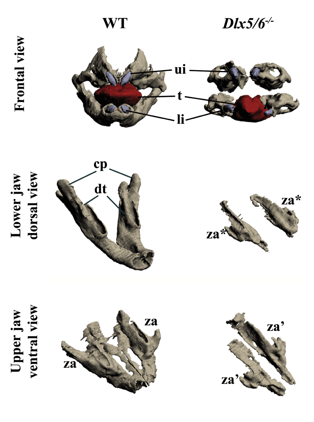
The skull of most vertebrates is characterized by the presence of articulated, asymmetric jaws which support the function of a muscularized oral cavity2,3. During embryonic development, the upper and lower jaws derive from the maxillary and mandibular processes of the first pharyngeal arch (PA1). Most cartilaginous and dermatocranial derivatives of PA1 are formed by Cranial Neural Crest Cells (CNCCs)49. During migration, signals emanating from the endoderm and possibly other PA1 components instruct the CNCCs to unfold the morphogenetic process of the jaws8,10,11. The nested expression of Dlx homeobox genes, vertebrate homologues of Drosophila Distal-less, has a fundamental role in the specification of the dorsoventral patterning of PA1 derivatives2,12. While Dlx1 and Dlx2 are expressed by CNCCs of the maxillary and mandibular components of PA1, Dlx5 and Dlx6 transcripts are present only in mandibular CNCCs. Targeted simultaneous inactivation of Dlx5 and Dlx613,14 results in the transformation of lower jaw into upper jaw-like structures, underlining the importance of these genes for lower jaw identity. The activation of Dlx5 and Dlx6 by endothelin-1 signalling is necessary and sufficient to define lower jaw identity1519. Interestingly it has been observed13,14 that, after inactivation of Dlx5 and Dlx6, maxillary components are also affected despite the fact that these genes are not expressed by maxillary CNCCs. This observation could be accounted for by the presence of shared Dlx5/6-dependent signalling centres in proximity to the extremities of both the mandibular and maxillary arches; this notion gave rise to the so-called “Hinge and Caps” model of jaw organization1,3,20. In its original formulation this model predicts the presence of two opposing morphogen gradients, one emanating from the region of the upper/lower jaw articulation (hinge) and one from the distal extremities of PA1 (caps); the origin and nature of these signals remain elusive. Here we revisit the effects of Dlx5 and Dlx6 double inactivation on jaw development and, using a transgenic lineage tracing approach, we reveal that the maxillary arch epithelium harbours a cellular contingent derived from frontonasal Dlx5-expressing progenitors. Our findings suggest that transient Dlx5/6 expression could program these epithelial cells to provide the cues needed for maxillary arch morphogenesis.

Material and methods

Mouse strains and breeding

All animal experimentation was performed in accordance to French national regulations and approved by the MNHN ethical committee (approval n° 68-028r1). For this study we used about 35 dams (including 10 WT, 5 Dlx5+/-; 3 Dlx5/6+/-; 12 B6.129S4-Gt(ROSA)26Sortm1Sor/J; 5 B6(Cg)-Dlx5tm1(cre/ERT2)Zjh/J) and analyzed about 120 embryos, the exact record of animals used, litters obtained, embryos genotyped and number of embryos per litter is on record in our animal house. WT animals were from Charles River France and were maintained in the MNHN mouse facility which is officially certified by the French National Animal well being committee.

Dlx5lacZ/+ knock-in mice were maintained on a mixed B6/D2 genetic background21. Double Dlx5 and Dlx6 (Dlx5/6) mutant mice were maintained and genotyped as reported22. The inducible Cre driver strain B6(Cg)-Dlx5tm1(cre/ERT2)Zjh/J (designed by Z. J. Huang23), and the Cre reporter strain B6.129S4-Gt(ROSA)26Sortm1Sor/J were purchased from Jackson Laboratory (#10705 and #003309 respectively; Maine, USA) through Charles River Laboratories (L’Arbresle, France) and maintained on a C57BL/6J genetic background through heterozygous mating. Double heterozygous embryos were obtained through bi-directional crosses. Induction of Cre recombinase activity was obtained upon single intraperitoneal injection of 5mg of tamoxifen (Sigma-Aldrich), in corn oil. Tamoxifen preparation and administration in pregnant dams followed the Jackson Laboratory’s Guidelines and CNRS/MNHN Animal Handling Guidelines. Dams were anesthetized in a chamber containing 2.5% isoflurane in oxygen and euthanized by cervical dislocation at indicated stages and embryos were collected in phosphate-buffered saline (PBS), then staged and fixed by immersion in ice-cold fixative (2% paraformaldehyde/0.2% glutaraldehyde) for 5 to 15 minutes (depending upon their developmental stage).

β-galactosidase detection

For lacZ expression, embryos were fixed for 15–30 min in 4% paraformaldehyde; X-gal staining was performed as described previously21,24. Vehicle (corn oil) injection in double heterozygous mice did not yield leaking ß-galactosidase activity.

Histology and 3D reconstruction

Heads from 18.5dpc (days post coitum) Dlx5/6-/- and wild type mouse embryos were fixed in Bouin’s solution (Sigma, France), embedded in paraffin and complete sets of frontal or parasagittal serial sections (12µm) were prepared. All sections were stained by Mallory’s trichrome as in19 and photographed (Nikon Digital Site DS-FI1). Pictures were aligned, piled and registered using the Fiji plug-in of NIH ImageJ “Register Virtual Stack Slices” (http://fiji.sc/wiki/index.php/Register_Virtual_Stack_Slices). 3D segmentation was performed with Mimics (Materialise, Belgium: http://biomedical.materialise.com/mimics) and visualized using Adobe Acrobat 9 pro.

Results

Dlx5/6 inactivation results in upper and lower jaw transformation with gain of symmetry

Previous reports suggest that double inactivation of Dlx5 and Dlx6 results in lower-to-upper jaw transformation; these reports also indicated that the upper jaw of these mice is not normal13,14. To better visualize the jaw phenotype of Dlx5/6 mutants, we performed 3D reconstructions of craniofacial elements of 18.5 dpc (days post coitum) embryos. Frontal view of the mutant jaws (Figure 1, upper panel) shows an obvious gain of symmetry compared to a WT animal. Examining the defects of the lower and upper jaws separately (Figure 1, middle and lower panels), it is evident that both are transformed. In the absence of Dlx5 and Dlx6 the dentary and the upper jaw bones do not form correctly and are replaced by remarkably similar skeletal structures. In the mutant embryos, both the upper and lower jaw skeletal elements are reduced in size, are not fused in the midline, and display a lateral process positionally homologous to the wild type zygomatic arch. Thus the upper and lower jaw mutant bones resemble each other more closely than usually found in their normal counterparts.

8f85a08a-a762-445a-b335-1dbd924b5b5a_figure1.gif

Figure 1. Three-dimensional reconstruction of the dentary and maxillary bones of 18.5dpc wild type and Dlx5/6-/- mouse embryos.

Upper row: Frontal view of WT and Dlx5/6-/- oral apparatus. Skeletal elements are grey, the tongue is red and incisors are violet. Middle row: Dorsal view of the dentary bone of WT and Dlx5/6-/- 18.5dpc mice. Lower row: Ventral view of the maxillary components of WT and Dlx5/6-/- 18.5dpc mice. Note that the inactivation of Dlx5/6 results in the transformation of both lower and upper jaw skeletal elements into new structures which appear more similar to each other than to their WT counterpart. cp, coronoid processes; dt, dentary bone; li, lower incisor; t, tongue; ui, upper incisor; za, zygomatic arch; za*, zygomatic arch-like structure deriving from lower jaw transformation; za’, zygomatic arch-like structure deriving from upper jaw transformation.

Transient Dlx5 expression in maxillary arch progenitors

In Dlx5-lacZ heterozygous Theiler stage (ts) 19 (12 dpc) embryos the reporter is active in the olfactory pit and mandibular arch, but not in the maxillary arch; this pattern of expression does not change upon tamoxifen treatment of the pregnant dam (Figure 2A,A’). To understand the origin of the Dlx5/6-dependent defect of the upper jaw we used a genetic approach to follow the lineage of Dlx5-precursors in the head. To this end we brought the R26R-lacZ reporter into the Dlx5-creERT2 driver background and we activated cre-recombinase activity by tamoxifen treatment of the pregnant dam at ts9 (7 dpc). We monitored ß-Gal reporter activity from ts15 (10 dpc) to ts20 (12.5 dpc). At ts15 we observed a stream of ß-Gal-positive cells extending from the lambdoidal junction, which joins the olfactory pit with the distal maxillary arch1,25, towards the body of the maxillary arch (Figure 2B,B’). At ts19 and ts20 (Figure 2C,C’; D,D’) reporter-expressing cells are found in the upper epithelial lining of the maxillary arch (arrowheads in Figure 2C’, 2D’) and in two distinct proximal and distal territories of the arch body (red asterisk in Figure 2C’).

8f85a08a-a762-445a-b335-1dbd924b5b5a_figure2.gif

Figure 2. Lineage of Dlx5-expressing cells in the maxillary arch.

ß-Galactosidase activity in the cephalic region of Dlx5-lacZ (A,A’) and Dlx5-creERT2; R26R-lacZ mouse embryos (BD’). In all cases pregnant dams were treated with tamoxifen at 7dpc/Theiler stage 9 (ts9) and embryos were collected at the indicated Theiler stage. (A,A’) As expected, even after tamoxifen treatment, Dlx5 is expressed in the mandibular arch (md), in the olfactory pit (olf), in the otic vesicle (ov), in the striatum (st) and in the hind limb (hl), but not in the maxillary arch. (B,B’) Permanent activation of lacZ reporter expression in derivatives of Dlx5-expressing early progenitors (ts9) reveals the presence of a positive cellular contingent in the ts15 lambdoidal junction (λ) between the olfactory pit and the maxillary process. (C,C’; D,D’) At later developmental stages (ts19, ts20) a contingent of lacZ positive cells populates the distal domain of the maxillary arch. hl, hind limb; md, mandibular arch; mx, maxillary arch; olf, olfactory pit; ov, otic vesicle; bt, basal telencephalon; λlambdoidal junction; red asterisk/black arrowheads, territories of the maxillary arch colonized by derivatives of Dlx5-expressing progenitors. Bar: AD 1mm; A’D’ 250µm.

To determine more precisely the tissue distribution of craniofacial derivatives of Dlx5-positive cells, we performed serial paraffin sections of Dlx5-creERT2; R26R-lacZ ß-Gal-stained mouse embryos (12.5 dpc) after tamoxifen treatment of pregnant dams at 7dpc/Theiler stage 9 (ts9) (Figure 3). While in the mandibular arch ß-Gal staining is limited, as expected, to CNCCs derivatives, only epithelial cells lining the maxillary arch are positive. As no Dlx5-positive epithelial cells are present in the maxillary arch of normal embryos, we conclude that a population of epithelial cells derived from the Dlx5-positive frontonasal process participates in the formation of the maxillary arch.

8f85a08a-a762-445a-b335-1dbd924b5b5a_figure3.gif

Figure 3. Derivatives of Dlx5-expressing progenitors in the upper and lower jaw primordia.

Sagittal section of Dlx5-creERT2; R26R-lacZ through the cephalic region of a 12.5 dpc/Theiler stage 22 (ts22) mouse embryo. The pregnant dam was treated with tamoxifen at 7dpc/ts9. ß-Galactosidase activity is found in CNCCs derivatives of the lower jaw (arrowheads). In contrast, in the upper jaw positive cells are only present in the overlying epithelium (arrows). mc, Meckel’s cartilage; asterisks, lower jaw muscles. Scale bar 50µm in insert, 150µm in the main figure.

Discussion

In this study we have re-examined the skeletal jaw phenotype of Dlx5/6 mutant mice. We confirm that both the mandibular and maxillary arches are transformed. The profound change in the shape of the maxillary arch is difficult to explain, as this region does not derive from a Dlx5/6-expressing territory. Indeed, in normal embryos maxillary CNCCs and the overlying epithelium do not express Dlx5/6. Lineage analysis to identify derivatives of Dlx5-positive progenitors reveals a new population of cells extending from the olfactory pit through the lambdoidal junction towards the maxillary arch1,20,25. These derivatives of Dlx5-positive cells have lost Dlx5 expression as seen by Dlx5 in situ hybridization (see for example14,21,26) and by lacZ-Dlx5 knock-in (21, and Figure 2A’). We have shown that early Dlx5 and Dlx6 expression in the anterior neural fold is essential for nasal capsule patterning27; our present findings suggest that the same population of cells could also contribute to maxillary patterning. This epithelial cell contingent might well exert a patterning role upon the maxillary arch providing spatial cues to the underlying mesenchyme. A further argument supporting the notion that Dlx5/6 patterning of the upper jaw does not require their expression in CNCCs derives from our recent observation that selective ablation of these genes in CNCCs does not affect upper jaw morphology (unpublished observation). This observation fits with the prediction of the ‘Hinge and Caps’ model1,3,20, and suggests that ‘cap’ signals could originate from derivatives of Dlx5-expressing frontonasal progenitors. Even if, after migration in the maxillary arch, these cells lose Dlx5 expression, it appears that the early expression of Dlx5 confers them the capacity to pattern maxillary arch CNCCs, which do not themselves express Dlx5 and Dlx6. It appears, therefore, that Dlx5 and Dlx6 pattern the upper and lower jaw through very different mechanisms, which must be coordinated to generate asymmetric, articulated, muscularized jaws.

Comments on this article Comments (0)

Version 3
VERSION 3 PUBLISHED 28 Nov 2013
Comment
Author details Author details
Competing interests
Grant information
Copyright
Download
 
Export To
metrics
Views Downloads
F1000Research - -
PubMed Central
Data from PMC are received and updated monthly.
- -
Citations
CITE
how to cite this article
Gitton Y, Narboux-Nême N and Levi G. Transitory expression of Dlx5 and Dlx6 in maxillary arch epithelial precursors is essential for upper jaw morphogenesis [version 2; peer review: 1 approved, 1 approved with reservations]. F1000Research 2014, 2:261 (https://doi.org/10.12688/f1000research.2-261.v2)
NOTE: If applicable, it is important to ensure the information in square brackets after the title is included in all citations of this article.
track
receive updates on this article
Track an article to receive email alerts on any updates to this article.

Open Peer Review

Current Reviewer Status: ?
Key to Reviewer Statuses VIEW
ApprovedThe paper is scientifically sound in its current form and only minor, if any, improvements are suggested
Approved with reservations A number of small changes, sometimes more significant revisions are required to address specific details and improve the papers academic merit.
Not approvedFundamental flaws in the paper seriously undermine the findings and conclusions
Version 2
VERSION 2
PUBLISHED 16 May 2014
Revised
Views
21
Cite
Reviewer Report 04 Aug 2014
Robert Knight, Craniofacial Development and Stem Cell Biology, King's College London, Guy's Hospital, London, UK 
Approved with Reservations
VIEWS 21
This paper by Gitton et al. re-examines the 'Hinge and Caps' model posited by Michael Depew and adds a new piece of evidence supporting this theory. This model argues that changes to mandibular and maxillary skeletal elements in mice mutant for ... Continue reading
CITE
CITE
HOW TO CITE THIS REPORT
Knight R. Reviewer Report For: Transitory expression of Dlx5 and Dlx6 in maxillary arch epithelial precursors is essential for upper jaw morphogenesis [version 2; peer review: 1 approved, 1 approved with reservations]. F1000Research 2014, 2:261 (https://doi.org/10.5256/f1000research.4412.r5678)
NOTE: it is important to ensure the information in square brackets after the title is included in all citations of this article.
  • Author Response 10 Sep 2014
    Giovanni Levi, Evolution des Régulations Endocriniennes, CNRS, UMR7221, Muséum National d'Histoire Naturelle, Paris, France
    10 Sep 2014
    Author Response
    “The authors display 3D reconstructions emphasising the symmetry of the transformed skeletal elements, but do not elaborate beyond the characterisations of these mutants that haven previously been performed.”
     
    As discussed in ... Continue reading
COMMENTS ON THIS REPORT
  • Author Response 10 Sep 2014
    Giovanni Levi, Evolution des Régulations Endocriniennes, CNRS, UMR7221, Muséum National d'Histoire Naturelle, Paris, France
    10 Sep 2014
    Author Response
    “The authors display 3D reconstructions emphasising the symmetry of the transformed skeletal elements, but do not elaborate beyond the characterisations of these mutants that haven previously been performed.”
     
    As discussed in ... Continue reading
Views
18
Cite
Reviewer Report 02 Jun 2014
Jennifer Fish, Orthopaedic Surgery, University of California, San Francisco, San Francisco, CA, USA 
Approved
VIEWS 18
The changes made by Gitton and colleagues in this revised manuscript have improved and clarified their new finding regarding Dlx5/6 contributions to upper jaw morphogenesis. In particular, the addition of Figure 3 and the changes to the text more clearly ... Continue reading
CITE
CITE
HOW TO CITE THIS REPORT
Fish J. Reviewer Report For: Transitory expression of Dlx5 and Dlx6 in maxillary arch epithelial precursors is essential for upper jaw morphogenesis [version 2; peer review: 1 approved, 1 approved with reservations]. F1000Research 2014, 2:261 (https://doi.org/10.5256/f1000research.4412.r4818)
NOTE: it is important to ensure the information in square brackets after the title is included in all citations of this article.
Version 1
VERSION 1
PUBLISHED 28 Nov 2013
Views
45
Cite
Reviewer Report 10 Feb 2014
Jennifer Fish, Orthopaedic Surgery, University of California, San Francisco, San Francisco, CA, USA 
Approved with Reservations
VIEWS 45
In this manuscript, Gitton and colleagues explore the role of Dlx5/6 in upper jaw morphogenesis. Dlx5/6 have largely been recognized for their role in lower jaw identity, based on the fact that loss of these genes in mice results in a ... Continue reading
CITE
CITE
HOW TO CITE THIS REPORT
Fish J. Reviewer Report For: Transitory expression of Dlx5 and Dlx6 in maxillary arch epithelial precursors is essential for upper jaw morphogenesis [version 2; peer review: 1 approved, 1 approved with reservations]. F1000Research 2014, 2:261 (https://doi.org/10.5256/f1000research.3104.r3486)
NOTE: it is important to ensure the information in square brackets after the title is included in all citations of this article.
  • Author Response 06 May 2014
    Giovanni Levi, Evolution des Régulations Endocriniennes, CNRS, UMR7221, Muséum National d'Histoire Naturelle, Paris, France
    06 May 2014
    Author Response
    We want, first of all, to thank Dr. Fish for her rapid review of our report. Her suggestions gave us the possibility to modify and, in our view, to improve ... Continue reading
COMMENTS ON THIS REPORT
  • Author Response 06 May 2014
    Giovanni Levi, Evolution des Régulations Endocriniennes, CNRS, UMR7221, Muséum National d'Histoire Naturelle, Paris, France
    06 May 2014
    Author Response
    We want, first of all, to thank Dr. Fish for her rapid review of our report. Her suggestions gave us the possibility to modify and, in our view, to improve ... Continue reading

Comments on this article Comments (0)

Version 3
VERSION 3 PUBLISHED 28 Nov 2013
Comment
Alongside their report, reviewers assign a status to the article:
Approved - the paper is scientifically sound in its current form and only minor, if any, improvements are suggested
Approved with reservations - A number of small changes, sometimes more significant revisions are required to address specific details and improve the papers academic merit.
Not approved - fundamental flaws in the paper seriously undermine the findings and conclusions
Sign In
If you've forgotten your password, please enter your email address below and we'll send you instructions on how to reset your password.

The email address should be the one you originally registered with F1000.

Email address not valid, please try again

You registered with F1000 via Google, so we cannot reset your password.

To sign in, please click here.

If you still need help with your Google account password, please click here.

You registered with F1000 via Facebook, so we cannot reset your password.

To sign in, please click here.

If you still need help with your Facebook account password, please click here.

Code not correct, please try again
Email us for further assistance.
Server error, please try again.