Keywords
Malaria, Nigeria, Plasmodium falciparum, PfPR2-10
This article is included in the Emerging Diseases and Outbreaks gateway.
Malaria, Nigeria, Plasmodium falciparum, PfPR2-10
Malaria is caused by one of five currently known Plasmodium species causing diseases in humans. These are P. falciparum, P. vivax, P. ovale, P. malariae and P. knowlesi. Scientists have modelled the anopheles mosquito vector extensively based on its characteristics, but not so much the parasite1. An absence of the Duffy antigen on red blood cells of West Africans has long been postulated to be responsible for the absence of P. vivax in these areas, but cases of P. falciparum, P. malariae and P. ovale, in order of decreasing occurrence, have been found. The World Malaria Reports of 20142 and 20153 reported 100% of confirmed cases in Nigeria as being due to P. falciparum. It is generally thought to account for about 98% of all malaria cases, with P. malariae accounting for the rest, often as a co-infection with P. falciparum4. This figure likely over-estimates the proportion of cases as P. falciparum is responsible for most severe cases of malaria; these are the cases most commonly reported alongside confirmed cases of malaria, which are tracked by passive surveillance in Nigeria. It may also be because of limited expertise in identifying other species of Plasmodium.
Available data from across Nigeria shows a mixed picture; large areas across northern Nigeria in 1967/68 found an average proportion of 22% of malaria parasitaemia due to P. malariae in a study at a time when most of Nigeria was considered holoendemic for malaria. P. malariae was often seen in coinfection with P. falciparum, particularly among younger age groups. P. ovale was responsible for 5% of malaria infections, being more common in children under five years of age, and P.falciparum ranged between 84.4% and 90.5% across age groups5. The proportion of P. malariae was high, probably because the data was obtained from a survey of both asymptomatic and symptomatic participants.
More recently, in 2010, prior to the commencement of nationwide LLIN distribution, a study including 4209 individuals in Jos, northern Nigeria, found a P. malariae rate as low as 1.6%, with P. falciparum responsible for 98.7% of infections, sometimes in co-infection with P. malariae4. Children aged less than 10 years and all individuals in every third household were selected for this abridged malaria indicator survey (MIS). Crucially, however, no P. vivax or P. ovale species were seen either in this location or another in south-eastern Nigeria, which was shown to have a higher rate of P. malariae infections (around 30%) and lower rate of P. falciparum infections (68.1%) in a study also conducted in 20106. This study, however, included 2,936 individuals from 1400 clusters in Abia state, spread out across the state. Quality control measures in the identification of parasite species were implemented in the study, including a WHO-certified malaria microscopist, giving credibility to the results obtained6. In south-western Nigeria, a study in Ikorodu in 2012 recruited 1,496 participants of all ages, which included 237 children under the age of five years and 509 children aged 15 years and below. Microscopy and DNA evaluation was used to determine parasite species, which found that 93.6% of participants had P. falciparum infection, with the remainder being P. malariae7. This is in contrast to previous studies in 1976 around the same location in south-western Nigeria, which found that 13% to 16% of parasitaemia was due to P. malariae, with 62 to 76% being due to P. falciparum8. These proportions were the age-group specific parasite prevalence in the study, which included mostly children aged five to 10 years (1,500 participants) in an attempt to compare spleen and parasite rates among individuals with sickle cell trait and those with normal adult haemoglobin. It also found P. ovale in 2% to 3% of participants in 5–10 and 2–4 year age groups, respectively. These findings, although limited in scope, perhaps suggest a changing trend, with the disappearance of P. ovale from Nigeria over time. P. falciparum is an undisputed leader in all the studies performed. Its high proportion perhaps accounts for high rates of malaria-related anaemia in most studies, explained by the ability of the parasite to invade and destroy both young and senescent red blood cells7,9.
We conducted this study to determine relative proportions of parasites causing clinical malaria in Sokoto, north-western Nigeria.
Ethical approval was obtained from the Independent Ethics committee of Usmanu Danfoidyo University Teaching Hospital, Sokoto, Nigeria with ethical approval number UDUTH/HREC/2014/No. 246. Ethical approval was also obtained from the Independent Ethical committee of the Sokoto State Ministry of Health with the approval number SMH/1580/VIV. Permission was also obtained from the district heads of the included communities in the study to visit the communities.
The study was conducted in Wamakko Local Government Area of Sokoto State, located in the north-western geopolitical zone of Nigeria. It has an area of 732.146km2, with a projected population of 260,860 by 201910. It is located at coordinates 13°2′16″N 5°5′37″E. The geography of the area is predominantly flat plains with Sudan Savannah-type vegetation and it stands at an altitude of 292m above sea level, near to the confluence of the Sokoto and Rima rivers. Its climate is tropical, described as local steppe climate.
The study was a two–point, cross-sectional prospective descriptive study conducted during the rainy season and dry season. In April and November 2016, we screened and recruited participants simultaneously until we reached the target population.
We determined the minimum sample size using Cochran’s formula11, assuming a prevalence of 50% based on a previous survey12, and 500 participants gave a power of at least 80% to show reliable results.
As is the norm for MIS’s, multistage cluster sampling in proportion to size was employed. The primary clusters were four randomly selected wards of the eleven political wards within the Local Government Area (LGA) based on the population within each of the wards, with secondary clusters being eight settlements unevenly selected from the four wards; proportionate to size, making up the total sample size. Based on the assumptions of a 70% response rate and that 80% of households include at least one child less than five years of age, in accordance with a previous Nigeria MIS in 201013, approximately 892 households were required to meet the target of at least 500 participants per season based on the assumptions stated earlier. This was surpassed by the estimated number of households within the eight settlements selected. All children in the secondary clusters who fulfilled the inclusion criteria, and whose parents consented to participate, were included in the study.
Participants were visited at their homes and while in the household, after identifying the household head. They were provided with information regarding the study and all eligible children within the household invited to participate. Those parents who accepted signed or thumb-printed the informed consent form. All children in the selected settlements who met the age criteria of two to 10 years, with or without symptoms of malaria, were recruited for the study, provided they had been residents of the study area for at least two weeks. They were, however, excluded if they were suspected to have taken any medication with antimalarial properties within the two weeks prior to enrolment36. Recruitment was carried out on consecutive days until the entire village was covered. Each participant was evaluated once except for those who had parasitaemia without symptoms, who were followed up by a field assistant for up to 48 hours for the development of symptoms. The period of recruitment was about a month in each season.
We conducted the study procedures at a central location in each of the study villages. Field assistants went from house-to-house and recruited the participants and then brought the consenting participants to the central location. A paediatrician screened potential participants for eligibility and the caregivers of eligible participants were required to sign informed consent forms. He performed a physical examination for each participant and graded splenomegaly according to Hackett’s criteria14. The WHO criteria for severe malaria was used15. Using a single use lancet, we collected capillary blood by pricking the index finger of the child’s left hand. A drop of blood was collected each for a thick and thin malaria parasite film for estimation of parasite density and species identification, respectively.
Concomitantly, during the same session, we did rapid diagnosis of malaria using a drop of blood with CareStart® Malaria HRP2 rapid detection tests (RDTs) (Access Bio, Inc., model G0141), which can detect P. falciparum.
Each day, we transported the samples to the paediatric department laboratory of the Usmanu Danfodiyo University Teaching Hospital and fixing of thick films was done with methanol. Thin films were stained immediately and stored in the lab. We analysed the samples in a completely anonymized manner in pairs of thick and thin films; examining the thin film if we found the thick film positive for malaria. The study numbers were the only identifiers for the thick films, which were kept apart from the thin films. A trained malaria microscopist performed the analysis, under the supervision of a medical parasitologist. We examined at least 10 fields before a slide was declared negative for malaria parasites.
The tail segment of the thin films was viewed to identify the species of malaria parasite, using the typical description of parasite species, having been trained on parasite identification16.
Quality control of the diagnosis of the parasitaemia was provided by a trained medical microbiologist re-examining 10% of the slides selected at random. A discrepancy of 10% or more would have necessitated reanalysis of all the thick films and the thin films subsequently. The discrepancy was 3% (kappa score of 0.71) and as such, this was not necessary.
Study participants with malaria, determined by the presence of at least one symptom and a positive RDT or thick blood film, were treated by the study paediatrician at home with Artemether-Lumefantrine. Children were dosed according to standard dosing17 but only the first dose was directly observed.
We analysed the data using SPSS version 22. We determined the prevalence of malaria by parasitaemia and RDT by determining proportions. We used descriptive statistics to determine averages and proportions. Participants with missing data were excluded from the analysis. We carried out sub-group analysis for age, gender and season and used kappa analysis to control the quality of malaria diagnosis.
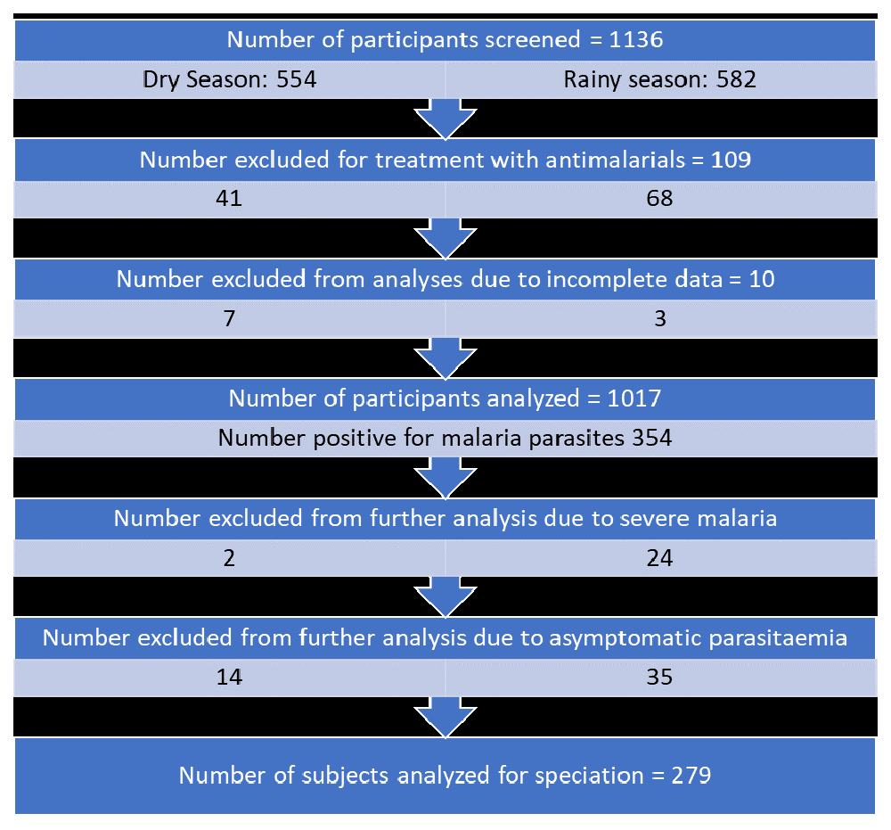
We screened a total of 1136 participants for inclusion in the study after they consented to participation in the study. We excluded 109 because they had been treated with antimalarials in the two weeks prior to enrolment and excluded 10 from the analysis due to incomplete data. We included 1017 participants in the analysis (Figure 1)18.
The age-sex distribution showed that all ages were equally represented in the study, as shown in Table 1.
We found an overall prevalence of malaria for the study of 34.8% using microscopy and 33.8% using RDT as shown in Table 2. There was an agreement between the two diagnostic methods, as shown by the kappa statistic (p <0.001).
We saw the highest age-specific prevalence among participants aged two years, with the lowest among ten-year olds. Table 3 also shows a significant association between the age of the participants and prevalence of malaria parasitaemia (p= 0.000).
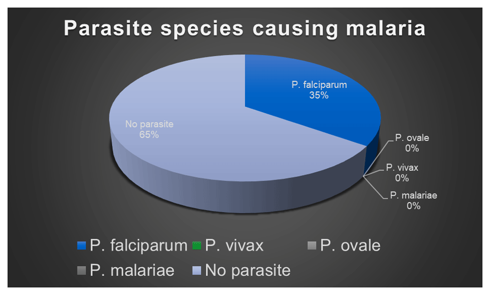
279 participants were found across the seasons to have clinical malaria and all of them had P. falciparum malaria, irrespective of the season and nature of their clinical presentation. The relative proportions of parasites are depicted in Figure 2. The most common presenting feature among these was fever (92%), followed by vomiting (38%), refusal to feed/poor appetite (32%) and body weakness (25%).
Of the clinical cases of malaria, 9.6% had complicated malaria, as indicated by the WHO criteria for severity19. The number of severe malaria cases was significantly lower in the dry season than the rainy season, as shown in Table 4. Different participants had various combinations of the criteria for severity, although the common criteria were hyperpyrexia, prostration and persistent vomiting.
The prevalence of malaria parasitaemia during the rainy season was significantly higher than the dry season, with prevalence rates of 49.3% and 20.2%, respectively, all due to P. falciparum.
The mean parasite density was much higher during the rainy season (1006.13) than during the dry season (405.45). The details are shown in Table 5.
The prevalence of malaria in this study, when compared with serial MIS’s performed in 201013 and 201520 shows a progressive reduction; from 48.1% to 37.1% for north-western Nigeria, and a prevalence of 46.6% for Sokoto in 2015 compared with 34.8% for our study. Prevalence in the MIS’s was measured among children aged six to 59 months and is probably higher than for the children included in this study because including the children from 6–10 years is likely to reduce the overall prevalence, as is excluding those aged six months to two years, who generally have a higher prevalence rate21.
Although there were studies performed in the past in Sokoto, they are limited in comparison to the present study by virtue of having been conducted in a different age group, or hospital in lieu of community setting and the seasons in which these studies were conducted. The prevalence of 34.8% found here was higher than the 27.9% found by Abdullahi et al.12 in Sokoto; however, samples in the previous study were collected from patients visiting two hospitals within the metropolis and was thus not community-based. Furthermore, all ages from 0 to 65 years were included in the study, which is likely to further dilute the findings and give a falsely low prevalence because the incidence of malaria is generally lower among adolescents and adults, as indicated in the study. The overall picture supports the suggestion of a reduction in the prevalence of malaria, likely owing to better access to malaria prevention and increasing urbanisation; both of which cause a decline in malaria parasite rates generally22.
The prevalence found in this study is also lower in comparison to the 45.4% prevalence rate found in a study by Jiya et al.23, conducted in Sokoto between 2007 and 2009. Additionally, it was lower than the prevalence of 49.6% found among children under the age of five years in the same study. Considering both age-specific prevalence rates, there is a reduction in prevalence, although being a hospital-based study, the prevalence for the former study is likely to be higher than the current. It is, however slightly, higher than the projected national average of 29% for 2015, with wide inter-regional differences24. The Nigerian MIS of 2015 found a higher prevalence of 46.6% than this study, although the age of included participants ranged from six to 59 months, which will limit the comparability of results from this study due to the different age ranges of participants20.
The prevalence by age in this study roughly indicated a progressive decline with age. The highest age-specific prevalence was among two-year-olds (50.4%), with a statistically significant difference among the age groups. This finding is in conformity with the steady-state assumption and is similar to findings in previous studies that showed higher prevalence among younger age-groups.
With respect to the parasite species causing uncomplicated malaria, all parasitaemia in this study was found to be due to P. falciparum. This is in keeping with recent studies in Adamawa25 and Cross River states9 in 2011 and 2013, respectively. Another study from Ihiala, in Anambra state of south-eastern Nigeria, found P. falciparum mono-parasitaemia even though this study considered all types of malaria, both severe and uncomplicated26. This is, however, unlikely to affect the findings, as most cases of severe malaria in this area are due to P. falciparum26. An earlier report from Sokoto between 2005 and 2006, carried out at Usmanu Danfodiyo University Teaching Hospital by Jiya and Sani23,27 likewise did not find any Plasmodium species apart from P. falciparum, although they only considered cases of severe malaria, which are unlikely to be due to a different species of Plasmodium within Nigeria. Meanwhile, only three cases out of 582 (0.01%) were positive for P.malariae in another study by Nwaorgu and Orajaka28 in Awka, south-eastern Nigeria. The finding of P. falciparum mono-parasitaemia supports the fact that P. falciparum is the dominant species of Plasmodium in Sub-Saharan Africa, with the tendency to exclude other forms of parasitaemia, as expounded by Lucas and Gilles29 in 1998 with time and sustained malaria control activities.
Other studies have shown the presence of other forms of parasitaemia, notably with P. malariae either as mono-infection or coinfection with P. falciparum. In Abia and Plateau states (2010), P. malariae accounted for 32.0% and 1.4% of malaria infections, respectively6. In a study in north-central Nigeria, 6.1% of examined participants had P. malariae infection30 and as high as 41% and 4%, respectively, had P. malariae and P. ovale infections in a historical study in Garki, Abuja (1968)31. Outside Nigeria, there has been a shift towards mono-parasitaemia with P. falciparum as well, and this has been documented in the Horn of Africa32 and Benin in West Africa33. Some authors have suggested it be an evidence of failing control measures but this is at variance with data from this study, which shows a reduction in prevalence from previous data, including a reduction in the number of severe cases of malaria, which were very few in this study.
Severe malaria was seen in 26 of the 1017 participants analysed in this study, with an overall prevalence of 2.6%. It was higher during the rainy than the dry season, probably due to higher prevalence of the disease and higher parasitaemia, as earlier discussed. This is lower than expected from other hospital-based studies for which children presenting to the hospital are more likely to be ill than those found in a community-based survey such as this. In one such study in Ilorin by Olanrewaju and Johnson29 found that a third of all children admitted with malaria had a severe form of malaria.
Figshare: complete data.xlsx. https://doi.org/10.6084/m9.figshare.11590542.v118.
Data are available under the terms of the Creative Commons Attribution 4.0 International license (CC-BY 4.0).
| Views | Downloads | |
|---|---|---|
| F1000Research | - | - |
|
PubMed Central
Data from PMC are received and updated monthly.
|
- | - |
Provide sufficient details of any financial or non-financial competing interests to enable users to assess whether your comments might lead a reasonable person to question your impartiality. Consider the following examples, but note that this is not an exhaustive list:
Sign up for content alerts and receive a weekly or monthly email with all newly published articles
Already registered? Sign in
The email address should be the one you originally registered with F1000.
You registered with F1000 via Google, so we cannot reset your password.
To sign in, please click here.
If you still need help with your Google account password, please click here.
You registered with F1000 via Facebook, so we cannot reset your password.
To sign in, please click here.
If you still need help with your Facebook account password, please click here.
If your email address is registered with us, we will email you instructions to reset your password.
If you think you should have received this email but it has not arrived, please check your spam filters and/or contact for further assistance.
Comments on this article Comments (0)