Keywords
Odontogenic carcinosarcoma, α-smooth muscle actin, vimentin, epithelial mesenchymal transition, Ki-67.
This article is included in the Oncology gateway.
Odontogenic carcinosarcoma, α-smooth muscle actin, vimentin, epithelial mesenchymal transition, Ki-67.
We changed the title because it didn’t follow the nomenclature stated in the new WHO classification.
We tried to improve the histopathological description of the lesion.
We edited the histopathological Figures 3 & 4 to clarify the features of the lesion.
We repeated the ki67 staining and measured the area percent by leica Qwin software Figure 6.
We contacted the surgeon to improve the follow-up information of the patient.
We tried to improve the discussion as recommended by the reviewer.
We wrote the take-home message in the conclusion section.
See the authors' detailed response to the review by João Paulo Silva Servato
See the authors' detailed response to the review by Oslei Paes de Almeida
Malignant odontogenic tumors are rare group of malignant neoplasms that arise from odontogenic remnants1. One of these neoplasms is odontogenic carcinosarcoma (OCS), an extremely rare mixed malignant odontogenic neoplasm in which both the epithelial and the ectomesenchymal components are cytologically malignant2. In the 1992 World Health Organization (WHO) classification of tumors, OCS was included in the malignant odontogenic tumors after Tanaka and co-workers (1991) first reported an odontogenic tumor with a mixture of malignant epithelial and ectomesenchymal components3. In the 2005 WHO classification, the tumor was removed due to an absence of current diagnostic criteria4. OCS has been added again in the 2017 edition because of the availability of cases with adequate diagnostic immunohistochemical and molecular criteria5,6. Owing to the scarcity of reported cases - only eleven reported cases in the English literature - OCS clinical behavior remains unexplored7,8. In the current work, we report a case of OCS with the detailed clinical, radiographic, histopathological and immunohistochemical description.
In December 2016, a 28-year-old Egyptian male patient working as an accountant was referred to the Department of Oral and Maxillofacial Surgery, Faculty of Dentistry, Cairo University, with a complaint of painless swelling of six months duration in the left side of his face measuring 7 × 6 cm. The patient had a pathological report of an incisional biopsy, performed outside our institute, which was diagnosed as ameloblastoma. There were no palpable lymph nodes. Intra-oral examination revealed an absence of the lower left molars with a fistula opening on the alveolar crest with no oozing pus.
A cone beam computed tomography examination revealed an ill-defined multilocular osteolytic lesion with fine radiopacities extending from the lower left second premolar up to the ramus. There was prominent cortical expansion with areas of perforation (Figure 1). A hemi-mandibulectomy was performed based on the incisional biopsy diagnosis and the pathological fracture of the mandible. The ramus was totally destructed and the tumor was invading the masseter muscle (Figure 2). Differential diagnosis was made based on the clinical examination, radiographic appearance and gross examination as: ghost cell odontogenic carcinoma, ameloblastic carcinoma, ameloblastic fibrosarcoma, ameloblastic carcinosarcoma, calcifying epithelial odontogenic tumor, atypical type of ameloblastic fibro-dentinoma9 and ameloblastoma.
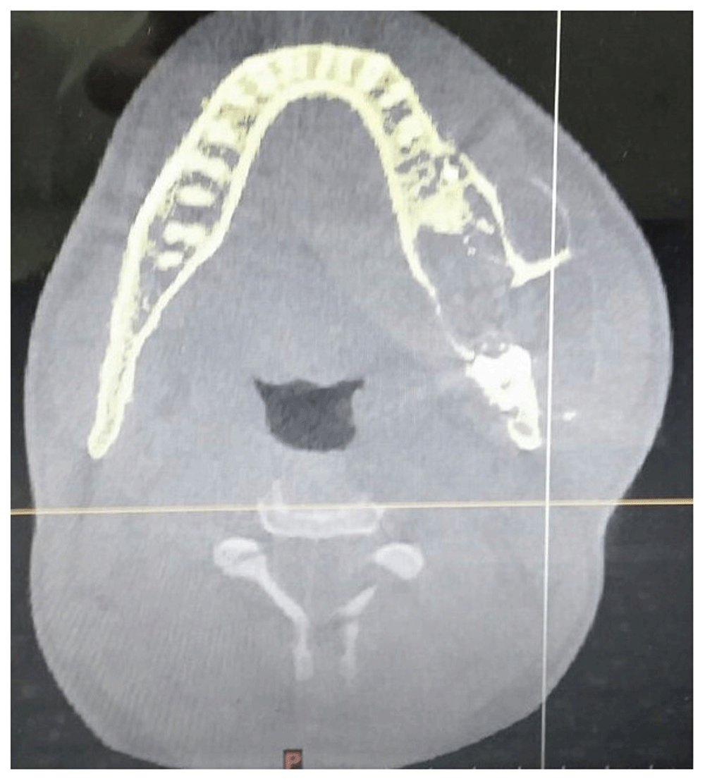
There was scattered radiopacities closely related to the impacted first molar. The second molar was displaced against the angle of the mandible.
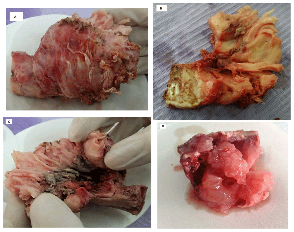
Gross examination: (A) Hemi-mandibulectomy with safety margin including the masseter muscle. (B) Cross-section of the resected specimen showing the fleshy neoplasm and the impacted first molar. (C) Top view of the resected specimen showing the necrotic fistula on the alveolar ridge. (D) The excisional specimen of the recurred lesion.
Histopathological examination revealed hypercellular follicles and strands of odontogenic epithelium, that were lined peripherally by multilayers of tall columnar ameloblast like cells. The center of the follicles was filled by stellate reticulum like cells, which were basaloid in appearance in some follicles. In between the follicles, there was a highly cellular primitive ectomesenchyme resembling dental papilla. Some of the nuclei of the ectomesenchymal cells were hyperchromatic. In some fields, dentinoid matrix was detected around the epithelial strands (Figure 3A).Based on the histopathological examination and the case report of Giraddi and Garg 20129, and because it was before the publication of the new WHO classification of 2017, a diagnosis was made of ameloblastic fibro-dentinoma (atypical type).

(A) Microscopic image showing odontogenic epithelial follicles formed of ameloblast-like cells on the periphery and stellate-reticulum like cells on the center, which were surrounded by primitive ectomesenchyme resembling dental papilla. Dentinoid material was detected in contact with the epithelial follicles (black arrows). (B) Microscopic image showing another field which was hypercellular in both components with mild pleomorphism and hyperchromatism.
After nine months and after the publication of 2017 WHO classification, the patient returned with another large painless swelling in the left temporal and infra-temporal fossa of three months duration. There were no palpable lymph nodes. An oral and maxillofacial surgeon used local anesthesia and an intra-oral approach to completely excise the recurrent lesion (Figure 2D). Augmentin 1gm tablet/12 hours for five days and anti-inflammatory medication were prescribed for the patient after the surgery. Histopathological examination revealed highly cellular neoplasm, which showed cellular atypia and increased nuclear cytoplasmic ratio in both epithelial and ectomesenchymal components. The typical ameloblastic architecture of the epithelial follicles was lost in some areas. The ectomesenchymal component showed large pleomorphic cells (Figure 4).
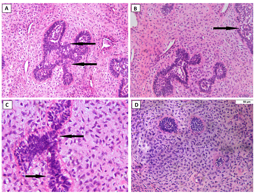
Histopathological examination of the recurred lesion (hematoxylin and eosin stain): (A & B) Microscopic image showing hypercellular epithelial follicles with plump hyperchromatic and pleomorphic cells and absence of the stellate reticulum cells in the center, along with hypercellular ectomesenchyme, the basement membrane was vague in some areas (black arrows) (X200). (C) Microscopic image showing epithelial follicle with pleomorphism, hyperchromatism, and increased nuclear-cytoplasmic ratio. The basement membrane was lost in some areas (black arrows) (X400). (D) Microscopic image showing atypia in ectomesenchymal component with increased mitosis, bizarre shaped nuclei and increased nuclear/ cytoplasmic ratio (X400).
The epithelial nests showed strong membranous positivity with AE1/AE3, while vimentin stained the ectomesenchyme diffusely and the epithelial nests patchily. Alpha-smooth muscle actin (α-SMA) showed positivity in the endothelial cells as well as in scattered epithelial cells (Figure 5). Ki-67 index was measured by Leica Qwin software and was 7.46% in the epithelial component, while in the ectomesenchymal component it was 2.78% (Figure 6). Based on the histopathological and immunohistochemical findings, the case was diagnosed as OCS. In 2019 and after nearly a year and a half, the patient came to Oral & Maxillofacial Surgery department for the reconstruction surgery and the examination revealed neither a recurrence nor complications.
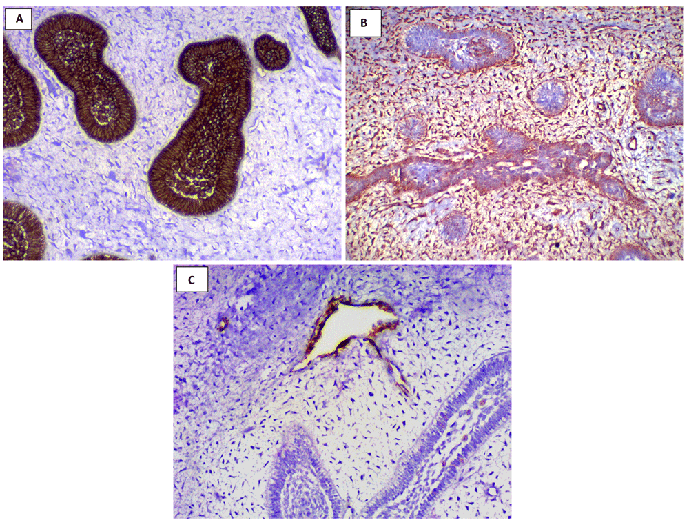
Immunohistochemical staining (X200): (A) Microscopic image showing strong membranous expression of AE1/AE3 in the epithelial component with the ectomesenchymal component being completely negative. (B) Microscopic image showing strong diffuse cytoplasmic expression of vimentin in ectomesenchymal component along with patchy expression in the epithelial component. (C) Microscopic image showing cytoplasmic expression of α-SMA in the endothelial cells and scattered epithelial cells.
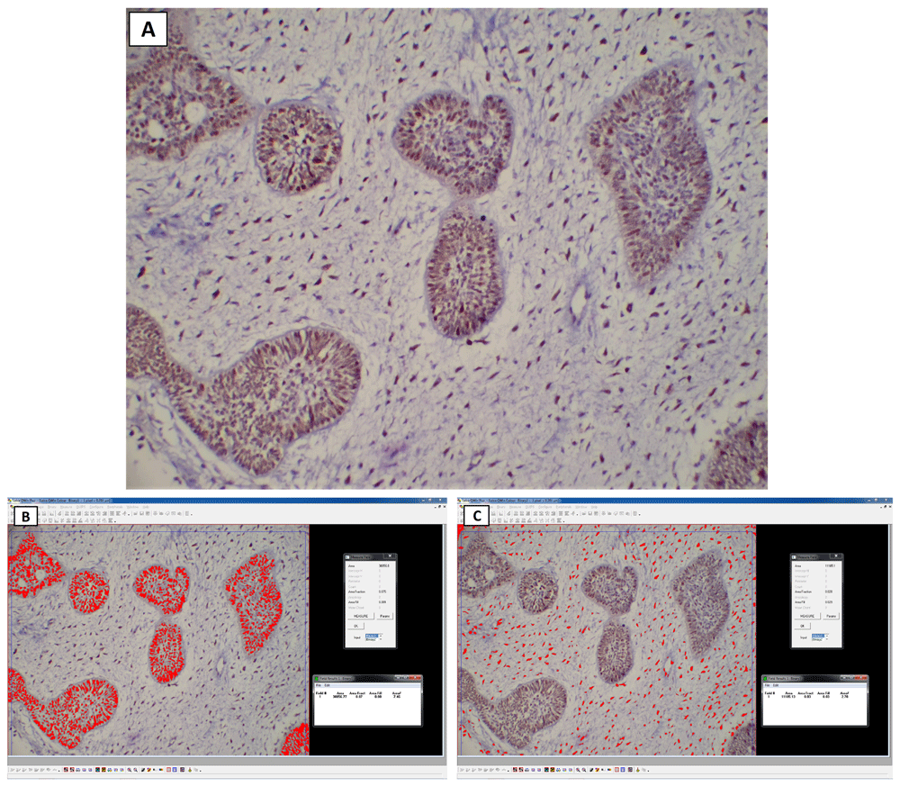
Immunohistochemical staining and analysis of Ki-67 (X200): (A) Microscopic image showing strong positive reaction of both epithelial and ectomesenchymal cells. (B) Screenshot showing the measurement of the area percent of Ki-67 expression of epithelial component using Leica Qwin software. (C) Screenshot showing the measurement of the area percent of Ki-67 expression of mesenchymal component.
OCS is a rare biphasic malignant odontogenic neoplasm that has the same architecture of ameloblastic fibroma, in which the epithelial and the ectomesenchymal components are cytologically malignant. It can develop de novo from odontogenic remnants or as a transformation from a preexisting odontogenic benign or malignant neoplasm6. This transformation may be attributed to multiple surgical procedures or recurrences of the neoplasm2. The present case arose from a preexisting immature odontoma which was formerly called ameloblastic fibrodentinoma. Kunkel et al. (2004), DeLair et al. (2007) and Chikosi et al. (2011) reported cases aroused from ameloblastic fibrosarcoma, ameloblastic fibroma and ameloblastoma, respectively2,10,11.
Our case was a 28-year-old male with the lesion affecting the posterior mandible. In the English literature, there was a male predilection and only one case that occurred in maxilla4. The most common radiographic picture of OCS is ill-defined multilocular radiolucency with cortical perforation6. The current case showed multiple radiopacities; may be due to the preexisting immature odontoma.
OCS must be distinguished from ameloblastic fibrosarcoma, in which the ectomesenchymal component only shows cellular atypia, and spindle cell variant of ameloblastic carcinoma, which lacks the ectomesenchymal component2–8. In the present case, there was a patchy positive expression of vimentin in the epithelial component. This could be explained as OCS undergoes epithelial mesenchymal transition, a process in which the polarized immotile epithelial cell changes to gain the mesenchymal phenotype and indicates more aggressive behavior of the neoplasm7,12,13.
In accordance with Dos Santos et al. (2018), α-SMA staining in the current case showed scattered staining in the epithelial cells4. Its expression in the epithelial cells could be attributed to the epithelial mesenchymal transition process; which increases the potentiality of the tumor cells to invade and metastasize14.
The proliferative index Ki-67 in our case was around 7.5% in the epithelial component and 3% in the ectomesenchymal component. This is in accordance with the work of Dos Santos et al. (2018) and Soares et al. (2019), who found that Ki-67 index was higher in the epithelial component than the ectomesenchymal component4–7.
Regarding the clinical behavior of OCS, it is a highly aggressive highly recurrent malignant neoplasm7. In our case, the patient encountered recurrence of the lesion after 9 months follow up with no evident metastasis. In the English literature, six out of the eleven cases showed recurrence of the lesion and only 4 cases showed metastasis; whether to lungs or lymph nodes6. Kunkel et al. (2004) reported the late metastasis of their case; which was evident about 5 years after the first diagnosis10. There were five out of the eleven cases encountered death6, one of them was actually due to systemic complication after the resection of the lesion7.
The principle line of treatment, as with other malignant odontogenic neoplasms, is surgical resection with a wide safety margin along with neck dissection. However, adjunctive radiotherapy is still a matter of question, and it may be helpful in cases with soft tissue invasion6.
OCS is an extremely rare odontogenic malignant neoplasm that shows very aggressive clinical behavior with multiple recurrences and possible metastasis. Close long follow-up is recommended; due to the possible late metastasis of the lesion. Immunohistochemical staining with vimentin, α-SMA and Ki-67 is helpful in the diagnosis of OCS.
All data underlying the results are available as part of the article and no additional source data are required.
Written informed consent for publication of their clinical details or clinical images was obtained from the patient.
| Views | Downloads | |
|---|---|---|
| F1000Research | - | - |
|
PubMed Central
Data from PMC are received and updated monthly.
|
- | - |
Provide sufficient details of any financial or non-financial competing interests to enable users to assess whether your comments might lead a reasonable person to question your impartiality. Consider the following examples, but note that this is not an exhaustive list:
Sign up for content alerts and receive a weekly or monthly email with all newly published articles
Already registered? Sign in
The email address should be the one you originally registered with F1000.
You registered with F1000 via Google, so we cannot reset your password.
To sign in, please click here.
If you still need help with your Google account password, please click here.
You registered with F1000 via Facebook, so we cannot reset your password.
To sign in, please click here.
If you still need help with your Facebook account password, please click here.
If your email address is registered with us, we will email you instructions to reset your password.
If you think you should have received this email but it has not arrived, please check your spam filters and/or contact for further assistance.
Comments on this article Comments (0)