Keywords
Fascin, SALL4, Ameloblastoma, Immunohistochemistry, Odontogenic cysts, Odontogenic tumors
This article is included in the Manipal Academy of Higher Education gateway.
Fascin, SALL4, Ameloblastoma, Immunohistochemistry, Odontogenic cysts, Odontogenic tumors
In this revised version of the manuscript we have defined the aims and objectives of the study. The introduction has been modified with elaborated data on the prevalence, characteristics of odontogenic tumors and cysts taken up for the study, recurrence rate of the odontogenic cysts and tumors and the association of the biomarkers (Fascin and SALL4) with these odontogenic lesions (Histopathological variants of ameloblastoma, Adenomatoid odontogenic tumor, Odontogenic keratocyst, Dentigerous cyst, Calcifying odontogenic cyst, Radicular cyst) has been discussed. The statistical reporting and data presentation have been put forward with description on variables and methods for each significance test performed also the tables have been modified with more clarity. The clinical details of the patients have been attached as a supplementary file and has been uploaded in the OSF repository.
See the authors' detailed response to the review by Konstantinos I Tosios
See the authors' detailed response to the review by Qi-Wen Man
See the authors' detailed response to the review by Pentti Nieminen
See the authors' detailed response to the review by Hope M Amm
See the authors' detailed response to the review by Sandhya Tamgadge
Odontogenic cysts and tumors are said to originate from odontogenic apparatus or oral epithelium. Ameloblastoma, the most common odontogenic tumor is known for its local but aggressive biological behaviour.1 The 2017 World Health Organisation (WHO) classification on ameloblastomas have reclassified them into Conventional, Unicyctic and Peripheral2 Literature review states among the odontogenic lesions, Ameloblastoma and Odontogenic keratocyst are locally aggressive and recurrent lesions, also the commonest and prevalent odontogenic tumor in Indian population is ameloblastoma which ranges from 14.02% to 71.4% when compared to other odontogenic tumors.3,4 The globally, pooled estimate of the incidence rate of ameloblastoma is 0.92 per million population per year.5 The recurrence varies among various populations, 9.8% according to a Chinese study,6 while in European multicenter study it is reported to be 19.3%,7also tumors larger than 6 cm and involving the soft tissues or adjacent anatomical structures are associated with early recurrence irrespective of method of surgery. Also conservatively (marsupialization, enucleation, curettage) treated cases have a high recurrence rate compared to radical treatment.6 However there is no concrete data pertaining recurrence on AOT, they are benign with rare recurrence. The other odontogenic cysts included were developmental viz DC and COC, wherein DC is associated with an impacted tooth while COC is associated with calcifications and ghost epithelial cells. RC an inflammatory odontogenic cyst is commonly associated with carious or non-vital tooth.2
Research to identify new markers to determine the biological behavior of odontogenic cysts and tumors is ongoing. Literature review reveals many preliminary observations with no concrete evidence of a single marker being specific to these tumors and hence there is a need to determine new markers.6 In this study, we have employed two markers: fascin and SALL4. Fascin, a 55-kDa is a cytoskeleton binding protein that bundle actin filaments, assists the cell in forming stress fibres (or ruffled borders or micro spikes) and assists cell motility and migration hence fascin can be used for predicting the aggressive clinical course of a tumor.7–10 Usually, in normal adult epithelial cells fascin expression is low or absent.11 The gene encoding fascin-1 in humans is located on chromosome 7. SALL4 is a stem cell marker and a master zinc-finger transcriptional factor, and a member of the spalt-like (SALL) gene family.12 SALL4 is mapped to chromosome 20q13.2 and plays its part in maintaining pluripotency and self-renewal of embryonic and hematopoietic stem cells by interacting with other molecules such as OCT4, SOX2 and NANOG.13–15 SALL4 incorporated along with OCT4, SOX2 and KLF4 (OSK) helps in forming stable induction of pluripotent cells (iPS) cells with a higher efficiency.16 Several studies noted the aberrant SALL4 expression in different types of malignant neoplasms and various autosomal dominant diseases such as Okihiro/Duane-radial ray syndrome, acro-renal-ocular syndrome, Instituto Venezolano de Investigaciones Cientificas syndrome (IVIC) and are suspected to cause thalidomide embryopathy.17–20 Literature review confirms fascin contributes for cell motility and migration in many studies (Pubmed 376 articles), SALL4 contributes to stemness along with other stem cell markers (SOX2, OCT4, and NANOG).8–13 Studies have shown the expression of these stem cell markers in Ameloblastoma & OKC except SALL4.14 Most of the studies in SALL4 are related to malignant soft tissue tumors,15,16 no reports are available of SALL4 expression in odontogenic lesions. SALL4 is activated by various pathways such as Wnt/β-catenin,15 PI3K/AKT, signalling pathway through targeting PTEN16 or Notch signalling pathway17 thus facilitating migration, invasion and proliferation, while Fascin is activated via PI3K/ Akt pathway.18Also literature reports cross talk between Wnt/β-catenin and PI3K/Akt pathways or simultaneous activation of these pathways contributing for proliferation and cell migration.19–21 Hence the present study was done to evaluate the expression of these two biomarkers in various odontogenic tumors (Histopathological variants of Ameloblastoma, AOT) and odontogenic cysts (OKC,DC,RC,COC).
Formalin fixed paraffin embedded tissue (FFPE) sections were retrieved from the Department of Oral and Maxillofacial Pathology, Manipal College of Dental Sciences, Manipal, India after obtaining approval from Institutional Ethical Committee, (IEC approval number 360/2019, IEC 156/2014). The samples taken up for the study were from year 2012-2017 which included 40 cases of ameloblastoma with histopathological variants viz plexiform (no:11), follicular (no:12), Unicystic (no:12), desmoplastic (no.5),6 cases of AOT, 15 cases each of OKC, DC, RC and 5 cases of COC. The cases selected did comply to inclusion and exclusion criteria. All the samples taken for the current study were prior to the patient receiving any treatment, cases with recurrence were excluded. The diagnosis of the above said odontogenic cysts and tumors were based on clinical and histological features (using H&E staining) according to WHO guidelines.2
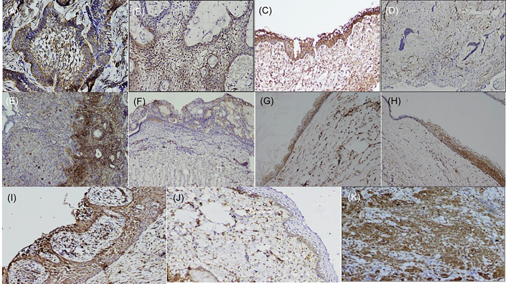
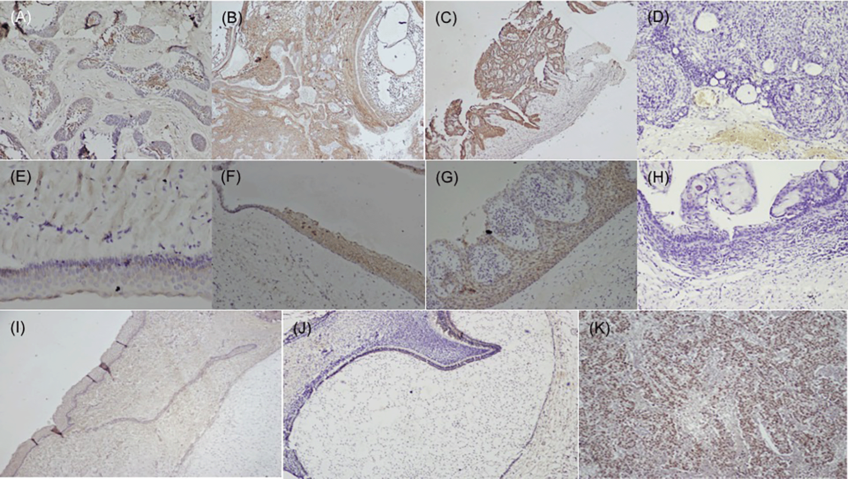
Immunohistochemical staining of the tissue sections from each of the cases selected was done using the streptavidin-biotin method. In brief, 4 μm sections were mounted on 3-aminopropyltriethoxysilane (APES) coated slides (Novolink Polymer Detection System, Novocastra). Sections were then deparaffinized in xylene, which was done in three grades for 10 minutes and hydrated in different grades of alcohol ranging from absolute alcohol (10 minutes), 95 % alcohol (10 minutes), 70% (10 minutes), 50% (10 minutes) each. Sections were then incubated with primary antibodies, rabbit antihuman SALL4 monoclonal antibody at a dilution of 1:100(IgG, clone EP-299, PathnSitu, Livermore, USA), mouse antihuman fascin monoclonal antibody (IgG1,clone 55K-2, SC-21743, Santa Cruz Biotechnology USA, Inc) diluted at 1:200. The sections were subsequently washed in tris-buffered saline and incubated with secondary biotinylated antibody and streptavidin-biotin peroxidase complex (Novolink Polymer Detection System, Novocastra) for 30 minutes each. Diaminobenzidine (DAB) was used as the chromogen and the sections were counterstained with Mayer’s hematoxylin. Buccal mucosa tissue was used as positive control,22 the basal cells of the epithelium were stained positive and endothelial cells within the lesional tissue were internal controls for fascin antibody (Figure 1), while dysgerminoma was taken as a positive control, for SALL4, for positive nuclear expression (Figure 2). Bud and bell stage of tooth development were also included for the study. The primary antibody was replaced during IHC staining for the negative control as per standard immunohistochemical protocol. The document of the protocol has been uploaded in the repository (Open Science Framework protocol.io).48

Histopathological variants of ameloblastoma: (A) Follicular (IHC, 10×), (B) Plexiform (IHC, 10×), (C) Unicystic (IHC, 10×), (D) Desmoplastic (IHC, 4×), (E) Focal immune-positivity for fascin in AOT (IHC, 10×), (F) COC (IHC, 10×), (G) OKC (IHC, 4×), (H) Dentigerous cyst (IHC, 10×), (I) Radicular cyst, (IHC, 10×), (J) Immuno-positivity for fascin in basal cells of the oral epithelium (IHC, 4×), (K) Oral squamous cell carcinoma used as positive control stained with fascin (IHC, 10×). IHC-Immunohistochemistry. The software used record images is Olympus-DP2BSW (ver 2.1).

Variants of ameloblastoma (A) Follicular (IHC, 10×), (B) Combination of follicular & plexiform (IHC,10×), (C) Unicystic (IHC, 4×), D) Immuno-negative in AOT (IHC, 10×), (E) OKC (IHC, 20×), (F) Dentigerous cyst (IHC, 10×), (G) Radicular cyst (IHC, 20×), (H) Immuno-negative COC (IHC, 10×), (I) Epithelial cells & ectomesenchyme surrounding the bud stage (IHC, 10×), (J) Bell stage: Focal positive to SALL4 in inner enamel epithelium (IEE) and sporadic expression in dental papilla (DP) (IHC, 10×), (K) Strong expression of SALL4 in dysgerminoma (positive control 20×). IHC-Immunohistochemistry.
Presence of brown color at the end of staining was considered as positive reactivity. The slides were evaluated with a light microscope (Olympus BX41) attached with Olympus DP20 microscope camera (Olympus Singapore Pvt Ltd, Singapore) at 20× & 40× magnification. The distribution of antibodies was assessed in the cytoplasm and cell membrane of ameloblastic lining of the lesions for fascin while SALL4 staining was evaluated in nuclear areas. In each case, three fields were randomly selected, and two observers independently evaluated the expression of these biomarkers, after selecting the most representative site separately under a light microscope at 200× and 400× magnification to eliminate the bias.
A semi-quantitative method was used to score the fascin and SALL4 expression in the epithelial odontogenic cells.
Based on intensity: (a) of the immunostaining in the epithelial odontogenic cells (0-1 = absent/weak, 2 = moderate, 3 = strong).
Degree of staining: (b) the percentage of positive odontogenic cells (1 ≤ 25% positive cells, 2 = 25-50% positive, 3 = 51-75% positive and 4 ≥ 75% positive cells).
Total staining: The final immunostaining score was determined by the sum of (a) + (b). Final scores ranged from 0 to 7 (0 = absent, 1-4 = weak and 5-7 = strong).
The data obtained was statistically analyzed with the statistical software program SPSS (version 17.0). The statistical significance of fascin and SALL4 in histopathological types of ameloblastoma was analysed using the chi-square test. P values less than 0.05 were considered to indicate statistical significance.
Immunohistochemically stained sections of various odontogenic cysts and tumors were evaluated for expression of fascin in the cell membrane, between cell boundaries and cytoplasm of peripheral ameloblastic cells, stellate reticulum like cells and stromal cells of 40 cases of ameloblastoma variants while expression of SALL4 was observed in the cytoplasm as well as nuclei of peripheral ameloblastic cells and stellate reticulum like cells. The total IRS score was the main outcome (Table 1, Table 2). The expression of fascin and SALL4 varied from case to case as well as in the same tissue section. Most of the variants of ameloblastoma were strongly positive for fascin but cases of desmoplastic ameloblastoma (5/5) were negative for fascin (Figure 1D). Fascin expression was found to be weak or absent in stellate reticulum like cells (Figure 1). In cases of unicystic ameloblastoma, positivity for fascin was observed in the basal as well as in the suprabasal layers (Figure 1C). However intra-group comparison did not show any significant difference. AOT was immune-positive to fascin in few areas (< 25%) with mild to moderate intensity (Figure 1E). Fascin expression in odontogenic cysts (OKC, RC, DC) (Figure 1G–I) was strongly positive with greater than 75% cells, while intensity ranged from moderate to strong along the cystic lining. COC revealed immune positivity ranging from 25-50% (Figure 1F). The SALL4 positivity was heterogeneous with varied intensity and staining pattern. In most of the histopathological variant of ameloblastoma, the immunopositivity observed, was diffuse in the cytoplasm and less localised to the nucleus (Figure 2A–C). The stromal cells were devoid of its expression except in the endothelial cells. SALL4 expression in odontogenic cysts was strongly positive with greater than 75% cells exhibiting diffuse cytoplasmic staining. Nuclear staining was evident in few cells (Figure 2E–G). COC was immune-negative (Figure 2H).
Regarding the evaluation of the statistical significance test, in all the odontogenic tumors, the staining intensity of fascin was similar compared to SALL4. With regard to the stained cell count, higher counts were observed with fascin as compared to SALL4. In relation to odontogenic cysts, OKC and DC, the intensity of fascin was more than SALL4. Also the higher cell counts were observed in fascin as compared to SALL4 in odontogenic keratocyst. The data regarding the same has been attached as Supplementary files(S1,S2,S3,S4) and has been uploaded in the repository (open science framework).
Researchers have worked on the molecular mechanism to understand the nature of local invasion of ameloblastomas into the surrounding tissues which include molecules degrading the extracellular matrix, those involved in bone remodelling, molecules associated with angiogenesis and molecules related to proliferation.23 Though the results are partially promising, the exact molecular mechanism of invasion in ameloblastomas is not completely understood.23–27 Cell motility is essential for tumor invasion and subsequent dissemination or metastases. This increase in motility occurs via the modulation of actin filaments to form finger-like plasma membrane protrusions termed invadopodia. Numerous actin-binding proteins, including fascin, regulate such dynamic rearrangement of the actin cytoskeleton. Fascin, being one of the actin cross-linking proteins, localizes to filopodia at the leading edge of migratory cells by organising f-actin into well-ordered, tightly packed parallel bundles observed in vitro studies.28
Fascin overexpression is observed in various precancerous lesions and oral squamous cell carcinoma (OSCC).29–33 In our study, we observed that a majority of our cases were strongly positive for fascin in the various subtypes of ameloblastoma. Various in vitro and in vivo studies have observed that fascin has a functional role in cell invasion and motility.34 This could account to the local aggressiveness of ameloblastoma clinically. Few of the ameloblastic follicles did not exhibit fascin, we speculate this could be attributed to loss of antigen during processing or reduced motility in these cells. Fascin expression in various cysts such as DC,OKC,RC and COC could be related to its influence in focal adhesion and cell dynamics.35
In various histopathological grades of ameloblastoma, SALL4 was expressed in the majority of cases. Studies have documented transcription activity of SALL4, which could be reflected by its positivity in the nucleus.36–44 We observed that the odontogenic epithelial cells were positive for SALL4 in the cytoplasm, stained diffusely, which we speculate could be in an inactive/dormant or mutant form which requires further investigation. Majority of OKC were devoid of SALL4 except in the basal cells. Radicular and dentigerous cysts, having marked infiltration of inflammatory cells had strong immune-positivity for SALL4 in the cytoplasm, an interesting finding of this study. Hence the role of cytokines in stimulating SALL4 needs to be ruled out. Odontogenic tumors, AOT and developmental odontogenic cysts, COC (simple type) were negative for SALL4. Studies have shown that OKCs expressed higher amount of PCNA and Ki-67 when compared to other jaw cysts, indicating its inherently increased proliferative potential of OKC.45 This speculates that various other molecular pathways could play an important role in the disease process. Further studies are required to explore this possibility, since this is a preliminary study.
Normal connective tissue cells such as fibroblasts, vascular endothelial cells, neural and glial cells, brain and splenic tissue expressed fascin, which relates to its function, required to maintain normal homeostasis.30 In embryogenesis, various migratory cells express fascin, except in terminally differentiated squamous cells where its expression is low or absent.30,46 Our previous study on tooth buds showed fascin expression in various stages of tooth development was site and time specific, thus confirming its role in cell remodulation.3 SALL4 expression was not detected in tooth bud stage, however focal positivity was observed in the cytoplasm of the epithelial cells of bell stage, this could attribute to the cells to undergo more differentiated state of the cells.47 The papillary cells in various stages of tooth germ were positive (Figure 2J) and this could relate to stemness due to the pluripotency nature of dental papilla. Further studies are required to understand the crosstalk with other stem cell markers in maintaining the stemness or pluripotency state of the cells.
In conclusion, the findings of the present study on the expression of fascin elucidate their role in motility and localized invasion or in maintaining the cellular homeostasis, while the expression of SALL4 remains elusive.
Open Science Framework: Expression of fascin and SALL4 in odontogenic cysts and tumors: an immunohistochemical appraisal. https://doi.org/10.17605/OSF.IO/9ZFRS.48
Data are available under the terms of the Creative Commons Zero “No rights reserved” data waiver (CC0 1.0 Public domain dedication).
| Views | Downloads | |
|---|---|---|
| F1000Research | - | - |
|
PubMed Central
Data from PMC are received and updated monthly.
|
- | - |
Competing Interests: No competing interests were disclosed.
Reviewer Expertise: Statistical reporting and data presentation
Is the work clearly and accurately presented and does it cite the current literature?
No
Is the study design appropriate and is the work technically sound?
Yes
Are sufficient details of methods and analysis provided to allow replication by others?
Partly
If applicable, is the statistical analysis and its interpretation appropriate?
Partly
Are all the source data underlying the results available to ensure full reproducibility?
Partly
Are the conclusions drawn adequately supported by the results?
Partly
Competing Interests: No competing interests were disclosed.
Reviewer Expertise: Immunohistochemistry, RNA sequencing, Odontogenic tumors
Is the work clearly and accurately presented and does it cite the current literature?
Partly
Is the study design appropriate and is the work technically sound?
Partly
Are sufficient details of methods and analysis provided to allow replication by others?
Yes
If applicable, is the statistical analysis and its interpretation appropriate?
I cannot comment. A qualified statistician is required.
Are all the source data underlying the results available to ensure full reproducibility?
Yes
Are the conclusions drawn adequately supported by the results?
No
Competing Interests: No competing interests were disclosed.
Reviewer Expertise: Oral pathology
Is the work clearly and accurately presented and does it cite the current literature?
No
Is the study design appropriate and is the work technically sound?
No
Are sufficient details of methods and analysis provided to allow replication by others?
No
If applicable, is the statistical analysis and its interpretation appropriate?
No
Are all the source data underlying the results available to ensure full reproducibility?
Partly
Are the conclusions drawn adequately supported by the results?
Partly
Competing Interests: No competing interests were disclosed.
Reviewer Expertise: Statistical reporting and data presentation
Alongside their report, reviewers assign a status to the article:
| Invited Reviewers | |||||
|---|---|---|---|---|---|
| 1 | 2 | 3 | 4 | 5 | |
|
Version 5 (revision) 27 Jun 24 |
read | ||||
|
Version 4 (revision) 10 Jun 24 |
read | read | read | ||
|
Version 3 (revision) 14 Dec 23 |
read | ||||
|
Version 2 (revision) 01 Sep 23 |
read | ||||
|
Version 1 23 Dec 22 |
read | read | read | ||
Provide sufficient details of any financial or non-financial competing interests to enable users to assess whether your comments might lead a reasonable person to question your impartiality. Consider the following examples, but note that this is not an exhaustive list:
Sign up for content alerts and receive a weekly or monthly email with all newly published articles
Already registered? Sign in
The email address should be the one you originally registered with F1000.
You registered with F1000 via Google, so we cannot reset your password.
To sign in, please click here.
If you still need help with your Google account password, please click here.
You registered with F1000 via Facebook, so we cannot reset your password.
To sign in, please click here.
If you still need help with your Facebook account password, please click here.
If your email address is registered with us, we will email you instructions to reset your password.
If you think you should have received this email but it has not arrived, please check your spam filters and/or contact for further assistance.
Comments on this article Comments (0)